Crystal Structures Reveal an Induced-fit Binding of a Substrate-like Aza-peptide Epoxide to SARS Coronavirus Main Peptidase.
Lee, T.W., Cherney, M.M., Liu, J., James, K.E., Powers, J.C., Eltis, L.D., James, M.N.G.(2007) J Mol Biology 366: 916-932
- PubMed: 17196984 Search on PubMedSearch on PubMed Central
- DOI: https://doi.org/10.1016/j.jmb.2006.11.078
- Primary Citation Related Structures:
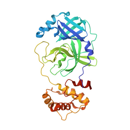
2GT7, 2GT8, 2GTB - PubMed Abstract:
The SARS coronavirus main peptidase (SARS-CoV M(pro)) plays an essential role in the life-cycle of the virus and is a primary target for the development of anti-SARS agents. Here, we report the crystal structure of M(pro) at a resolution of 1.82 Angstroms, in space group P2(1) at pH 6.0. In contrast to the previously reported structure of M(pro) in the same space group at the same pH, the active sites and the S1 specificity pockets of both protomers in the structure of M(pro) reported here are in the catalytically competent conformation, suggesting their conformational flexibility. We report two crystal structures of M(pro) having an additional Ala at the N terminus of each protomer (M(+A(-1))(pro)), both at a resolution of 2.00 Angstroms, in space group P4(3)2(1)2: one unbound and one bound by a substrate-like aza-peptide epoxide (APE). In the unbound form, the active sites and the S1 specificity pockets of both protomers of M(+A(-1))(pro) are observed in a collapsed (catalytically incompetent) conformation; whereas they are in an open (catalytically competent) conformation in the APE-bound form. The observed conformational flexibility of the active sites and the S1 specificity pockets suggests that these parts of M(pro) exist in dynamic equilibrium. The structural data further suggest that the binding of APE to M(pro) follows an induced-fit model. The substrate likely also binds in an induced-fit manner in a process that may help drive the catalytic cycle.
- Group in Protein Structure and Function, Department of Biochemistry, University of Alberta, Edmonton, Alberta, Canada.
Organizational Affiliation: