2GRO

Crystal Structure of human RanGAP1-Ubc9-N85Q


Experimental Data Snapshot

  • Method: X-RAY DIFFRACTION
  • Resolution: 1.70 Å
  • R-Value Free: 0.202 
  • R-Value Work: 0.185 

wwPDB Validation   3D Report Full Report


This is version 1.5 of the entry. See complete history


Literature

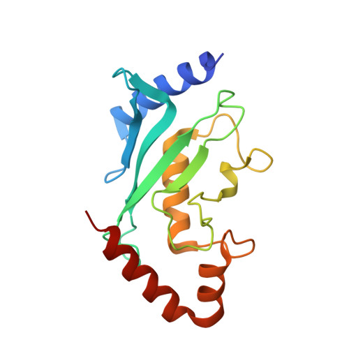
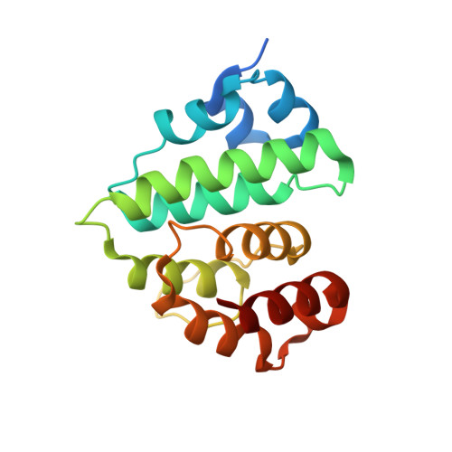
Lysine activation and functional analysis of E2-mediated conjugation in the SUMO pathway.

Yunus, A.A.Lima, C.D.

(2006) Nat Struct Mol Biol 13: 491-499

  • DOI: https://doi.org/10.1038/nsmb1104
  • Primary Citation of Related Structures:  
    2GRN, 2GRO, 2GRP, 2GRQ, 2GRR

  • PubMed Abstract: 

    E2 conjugating proteins that transfer ubiquitin and ubiquitin-like modifiers to substrate lysine residues must first activate the lysine nucleophile for conjugation. Genetic complementation revealed three side chains of the E2 Ubc9 that were crucial for normal growth. Kinetic analysis revealed modest binding defects but substantially lowered catalytic rates for these mutant alleles with respect to wild-type Ubc9. X-ray structures for wild-type and mutant human Ubc9-RanGAP1 complexes showed partial loss of contacts to the substrate lysine in mutant complexes. Computational analysis predicted pK perturbations for the substrate lysine, and Ubc9 mutations weakened pK suppression through improper side chain coordination. Biochemical studies with p53, RanGAP1 and the Nup358/RanBP2 E3 were used to determine rate constants and pK values, confirming both structural and computational predictions. It seems that Ubc9 uses an indirect mechanism to activate lysine for conjugation that may be conserved among E2 family members.


  • Organizational Affiliation

    Structural Biology Program, Sloan-Kettering Institute, New York, New York 10021, USA.


Macromolecules
Find similar proteins by:  (by identity cutoff)  |  3D Structure
Entity ID: 1
MoleculeChains Sequence LengthOrganismDetailsImage
Ubiquitin-conjugating enzyme E2 I161Homo sapiensMutation(s): 1 
Gene Names: UBE2IUBC9UBCE9
EC: 6.3.2.19
UniProt & NIH Common Fund Data Resources
Find proteins for P63279 (Homo sapiens)
Explore P63279 
Go to UniProtKB:  P63279
PHAROS:  P63279
GTEx:  ENSG00000103275 
Entity Groups  
Sequence Clusters30% Identity50% Identity70% Identity90% Identity95% Identity100% Identity
UniProt GroupP63279
Sequence Annotations
Expand
  • Reference Sequence
Find similar proteins by:  (by identity cutoff)  |  3D Structure
Entity ID: 2
MoleculeChains Sequence LengthOrganismDetailsImage
Ran GTPase-activating protein 1170Homo sapiensMutation(s): 0 
Gene Names: RANGAP1
UniProt & NIH Common Fund Data Resources
Find proteins for P46060 (Homo sapiens)
Explore P46060 
Go to UniProtKB:  P46060
PHAROS:  P46060
GTEx:  ENSG00000100401 
Entity Groups  
Sequence Clusters30% Identity50% Identity70% Identity90% Identity95% Identity100% Identity
UniProt GroupP46060
Sequence Annotations
Expand
  • Reference Sequence
Experimental Data & Validation

Experimental Data

  • Method: X-RAY DIFFRACTION
  • Resolution: 1.70 Å
  • R-Value Free: 0.202 
  • R-Value Work: 0.185 
  • Space Group: P 1
Unit Cell:
Length ( Å )Angle ( ˚ )
a = 39.295α = 72.58
b = 44.785β = 72.06
c = 61.027γ = 75.45
Software Package:
Software NamePurpose
DENZOdata reduction
SCALEPACKdata scaling
CNSrefinement
PDB_EXTRACTdata extraction
MAR345data collection
CNSphasing

Structure Validation

View Full Validation Report


Entry History 

Deposition Data

Revision History  (Full details and data files)

  • Version 1.0: 2006-05-30
    Type: Initial release
  • Version 1.1: 2008-05-01
    Changes: Version format compliance
  • Version 1.2: 2011-07-13
    Changes: Version format compliance
  • Version 1.3: 2017-10-18
    Changes: Refinement description
  • Version 1.4: 2021-10-20
    Changes: Database references
  • Version 1.5: 2024-02-14
    Changes: Data collection