A structural basis for complement inhibition by Staphylococcus aureus.
Hammel, M., Sfyroera, G., Ricklin, D., Magotti, P., Lambris, J.D., Geisbrecht, B.V.(2007) Nat Immunol 8: 430-437
- PubMed: 17351618 Search on PubMed
- DOI: https://doi.org/10.1038/ni1450
- Primary Citation Related Structures:
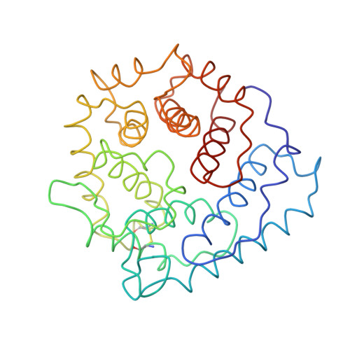
2GOM, 2GOX - PubMed Abstract:
To provide insight into bacterial suppression of complement-mediated immunity, we present here structures of a bacterial complement inhibitory protein, both free and bound to its complement target. The 1.25-A structure of the complement component C3-inhibitory domain of Staphylococcus aureus extracellular fibrinogen-binding protein (Efb-C) demonstrated a helical motif involved in complement regulation, whereas the 2.2-A structure of Efb-C bound to the C3d domain of human C3 allowed insight into the recognition of complement proteins by invading pathogens. Our structure-function studies provided evidence for a previously unrecognized mode of complement inhibition whereby Efb-C binds to native C3 and alters the solution conformation of C3 in a way that renders it unable to participate in successful 'downstream' activation of the complement response.
- Division of Cell Biology and Biophysics, School of Biological Sciences, University of Missouri at Kansas City, Kansas City, Missouri 64110, USA.
Organizational Affiliation: