2GJP | pdb_00002gjp

Structure of Bacillus halmapalus alpha-amylase, crystallized with the substrate analogue acarbose and maltose


Experimental Data Snapshot

  • Method: X-RAY DIFFRACTION
  • Resolution: 1.90 Å
  • R-Value Free: 
    0.214 (Depositor), 0.216 (DCC) 
  • R-Value Work: 
    0.172 (Depositor), 0.174 (DCC) 
  • R-Value Observed: 
    0.174 (Depositor) 

Starting Model: experimental
View more details

wwPDB Validation 3D Report Full Report

Validation slider image for 2GJP

Ligand Structure Quality Assessment 


This is version 2.1 of the entry. See complete history

Literature

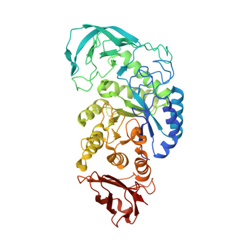
Structure of Bacillus halmapalus alpha-amylase crystallized with and without the substrate analogue acarbose and maltose.

Lyhne-Iversen, L.Hobley, T.J.Kaasgaard, S.G.Harris, P.

(2006) Acta Crystallogr Sect F Struct Biol Cryst Commun 62: 849-854

  • DOI: https://doi.org/10.1107/S174430910603096X
  • Primary Citation Related Structures: 
    2GJP, 2GJR

  • PubMed Abstract: 

    Recombinant Bacillus halmapalus alpha-amylase (BHA) was studied in two different crystal forms. The first crystal form was obtained by crystallization of BHA at room temperature in the presence of acarbose and maltose; data were collected at cryogenic temperature to a resolution of 1.9 A. It was found that the crystal belonged to space group P2(1)2(1)2(1), with unit-cell parameters a = 47.0, b = 73.5, c = 151.1 A. A maltose molecule was observed and found to bind to BHA and previous reports of the binding of a nonasaccharide were confirmed. The second crystal form was obtained by pH-induced crystallization of BHA in a MES-HEPES-boric acid buffer (MHB buffer) at 303 K; the solubility of BHA in MHB has a retrograde temperature dependency and crystallization of BHA was only possible by raising the temperature to at least 298 K. Data were collected at cryogenic temperature to a resolution of 2.0 A. The crystal belonged to space group P2(1)2(1)2(1), with unit-cell parameters a = 38.6, b = 59.0, c = 209.8 A. The structure was solved using molecular replacement. The maltose-binding site is described and the two structures are compared. No significant changes were seen in the structure upon binding of the substrates.


  • Organizational Affiliation
    • Department of Chemistry, Technical University of Denmark, Building 207, DK-2800 Kgs. Lyngby, Denmark.

Macromolecule Content 

  • Total Structure Weight: 58.03 kDa 
  • Atom Count: 4,302 
  • Modeled Residue Count: 481 
  • Deposited Residue Count: 485 
  • Unique protein chains: 1

Macromolecules

Find similar proteins by:|  3D Structure
Entity ID: 1
MoleculeChains  Sequence LengthOrganismDetailsImage
alpha-amylase485Sutcliffiella halmapalaMutation(s): 0 
EC: 3.2.1.1 (PDB Primary Data), 3.2.1.98 (UniProt)
UniProt
Find proteins for P19571 (Bacillus sp. (strain 707))
Explore P19571 
Go to UniProtKB:  P19571
Entity Groups
Sequence Clusters30% Identity50% Identity70% Identity90% Identity95% Identity100% Identity
UniProt GroupP19571
Sequence Annotations
Expand
Reference Sequence

Oligosaccharides

Help  
Entity ID: 2
MoleculeChains Length2D Diagram GlycosylationD Interactions
4,6-dideoxy-4-{[(1S,5R,6S)-3-formyl-5,6-dihydroxy-4-oxocyclohex-2-en-1-yl]amino}-alpha-D-xylo-hex-5-enopyranose-(1-4)-alpha-D-glucopyranose-(1-4)-beta-D-glucopyranose
B
3N/AN/A
Entity ID: 3
MoleculeChains Length2D Diagram GlycosylationD Interactions
4,6-dideoxy-4-{[(1S,5R,6S)-3-formyl-5,6-dihydroxy-4-oxocyclohex-2-en-1-yl]amino}-alpha-D-xylo-hex-5-enopyranose-(1-4)-alpha-D-glucopyranose-(1-4)-alpha-D-glucopyranose-(1-4)-alpha-D-glucopyranose
C
4N/AN/A
Entity ID: 4
MoleculeChains Length2D Diagram GlycosylationD Interactions
alpha-D-glucopyranose-(1-4)-alpha-D-glucopyranose
D
2N/A

Biologically Interesting Molecules (External Reference) 

1 Unique

Experimental Data & Validation

Experimental Data

  • Method: X-RAY DIFFRACTION
  • Resolution: 1.90 Å
  • R-Value Free:  0.214 (Depositor), 0.216 (DCC) 
  • R-Value Work:  0.172 (Depositor), 0.174 (DCC) 
  • R-Value Observed: 0.174 (Depositor) 
Space Group: P 21 21 21
Unit Cell:
Length ( Å )Angle ( ˚ )
a = 46.993α = 90
b = 73.497β = 90
c = 151.094γ = 90
Software Package:
Software NamePurpose
REFMACrefinement
ADSCdata collection
XDSdata scaling
MOLREPphasing

Structure Validation

View Full Validation Report


Ligand Structure Quality Assessment 


Entry History 

Deposition Data

Revision History  (Full details and data files)

  • Version 1.0: 2006-09-05
    Type: Initial release
  • Version 1.1: 2008-05-01
    Changes: Version format compliance
  • Version 1.2: 2011-07-13
    Changes: Version format compliance
  • Version 1.3: 2018-03-07
    Changes: Advisory, Data collection
  • Version 2.0: 2020-07-29
    Type: Remediation
    Reason: Carbohydrate remediation
    Changes: Advisory, Atomic model, Data collection, Derived calculations, Non-polymer description, Structure summary
  • Version 2.1: 2023-08-30
    Changes: Data collection, Database references, Refinement description, Structure summary