2GD8 | pdb_00002gd8

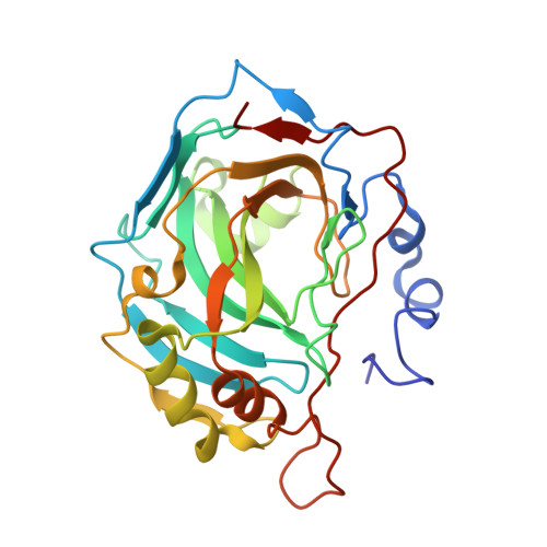
Crystal structure analysis of the human carbonic anhydrase II in complex with a 2-substituted estradiol bis-sulfamate


Experimental Data Snapshot

  • Method: X-RAY DIFFRACTION
  • Resolution: 1.46 Å
  • R-Value Free: 
    0.212 (Depositor), 0.208 (DCC) 
  • R-Value Work: 
    0.195 (Depositor), 0.190 (DCC) 

Starting Model: experimental
View more details

wwPDB Validation 3D Report Full Report

Validation slider image for 2GD8

Ligand Structure Quality Assessment 


This is version 1.4 of the entry. See complete history. 

Literature

2-substituted estradiol bis-sulfamates, multitargeted antitumor agents: synthesis, in vitro SAR, protein crystallography, and in vivo activity.

Leese, M.P., Leblond, B., Smith, A., Newman, S.P., Di Fiore, A., De Simone, G., Supuran, C.T., Purohit, A., Reed, M.J., Potter, B.V.

(2006) J Med Chem 49: 7683-7696

  • DOI: https://doi.org/10.1021/jm060705x
  • Primary Citation Related Structures: 
    2GD8

  • PubMed Abstract: 

    The anticancer activities and SARs of estradiol-17-O-sulfamates and estradiol 3,17-O,O-bis-sulfamates (E2bisMATEs) as steroid sulfatase (STS) inhibitors and antiproliferative agents are discussed. Estradiol 3,17-O,O-bis-sulfamates 20 and 21, in contrast to the 17-O-monosulfamate 11, proved to be excellent STS inhibitors. 2-Substituted E2bisMATEs 21 and 23 additionally exhibited potent antiproliferative activity with mean graph midpoint values of 18-87 nM in the NCI 60-cell-line panel. 21 Exhibited antiangiogenic in vitro and in vivo activity in an early-stage Lewis lung model, and 23 dosed p.o. caused marked growth inhibition in a nude mouse xenograft tumor model. Modeling studies suggest that the E2bisMATEs and 2-MeOE2 share a common mode of binding to tubulin, though COMPARE analysis of activity profiles was negative. 21 was cocrystallized with carbonic anhydrase II, and X-ray crystallography revealed unexpected coordination of the 17-O-sulfamate of 21 to the active site zinc and a probable additional lower affinity binding site. 2-Substituted E2bisMATEs are attractive candidates for further development as multitargeted anticancer agents.


  • Organizational Affiliation: 
    • Medicinal Chemistry, Department of Pharmacy and Pharmacology & Sterix Ltd., University of Bath, Bath BA2 7AY, United Kingdom.

Macromolecule Content 

  • Total Structure Weight: 30.5 kDa 
  • Atom Count: 2,471 
  • Modeled Residue Count: 257 
  • Deposited Residue Count: 259 
  • Unique protein chains: 1

Macromolecules

Find similar proteins by:|  3D Structure
Entity ID: 1
MoleculeChains  Sequence LengthOrganismDetailsImage
Carbonic anhydrase 2259Homo sapiensMutation(s): 0 
EC: 4.2.1.1 (PDB Primary Data), 4.2.1.69 (UniProt)
UniProt & NIH Common Fund Data Resources
Find proteins for P00918 (Homo sapiens)
Explore P00918 
Go to UniProtKB:  P00918
PHAROS:  P00918
GTEx:  ENSG00000104267 
Entity Groups
Sequence Clusters30% Identity50% Identity70% Identity90% Identity95% Identity100% Identity
UniProt GroupP00918
Sequence Annotations
Expand
Reference Sequence

Small Molecules

Ligands 4 Unique
IDChains Name / Formula / InChI Key2D Diagram3D Interactions
PO1

Query on PO1


Download:Ideal Coordinates CCD File
D [auth A],
E [auth A]
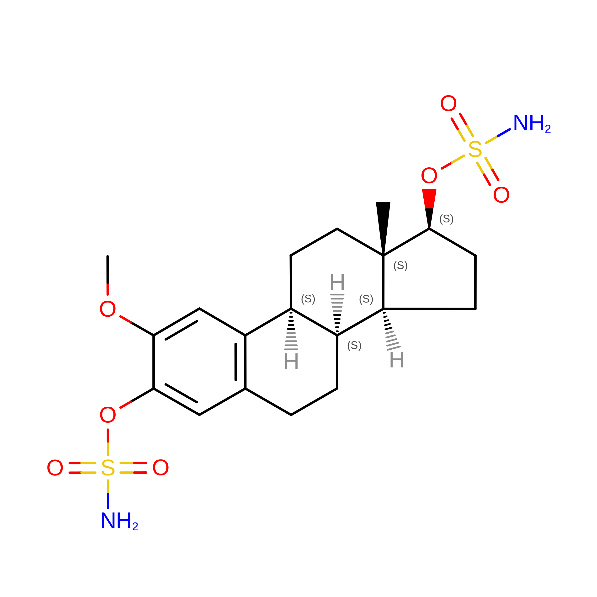
(9BETA,13ALPHA,14BETA,17ALPHA)-2-METHOXYESTRA-1,3,5(10)-TRIENE-3,17-DIYL DISULFAMATE
C19 H28 N2 O7 S2
AQSNIXKAKUZPSI-SSTWWWIQSA-N
MBO

Query on MBO


Download:Ideal Coordinates CCD File
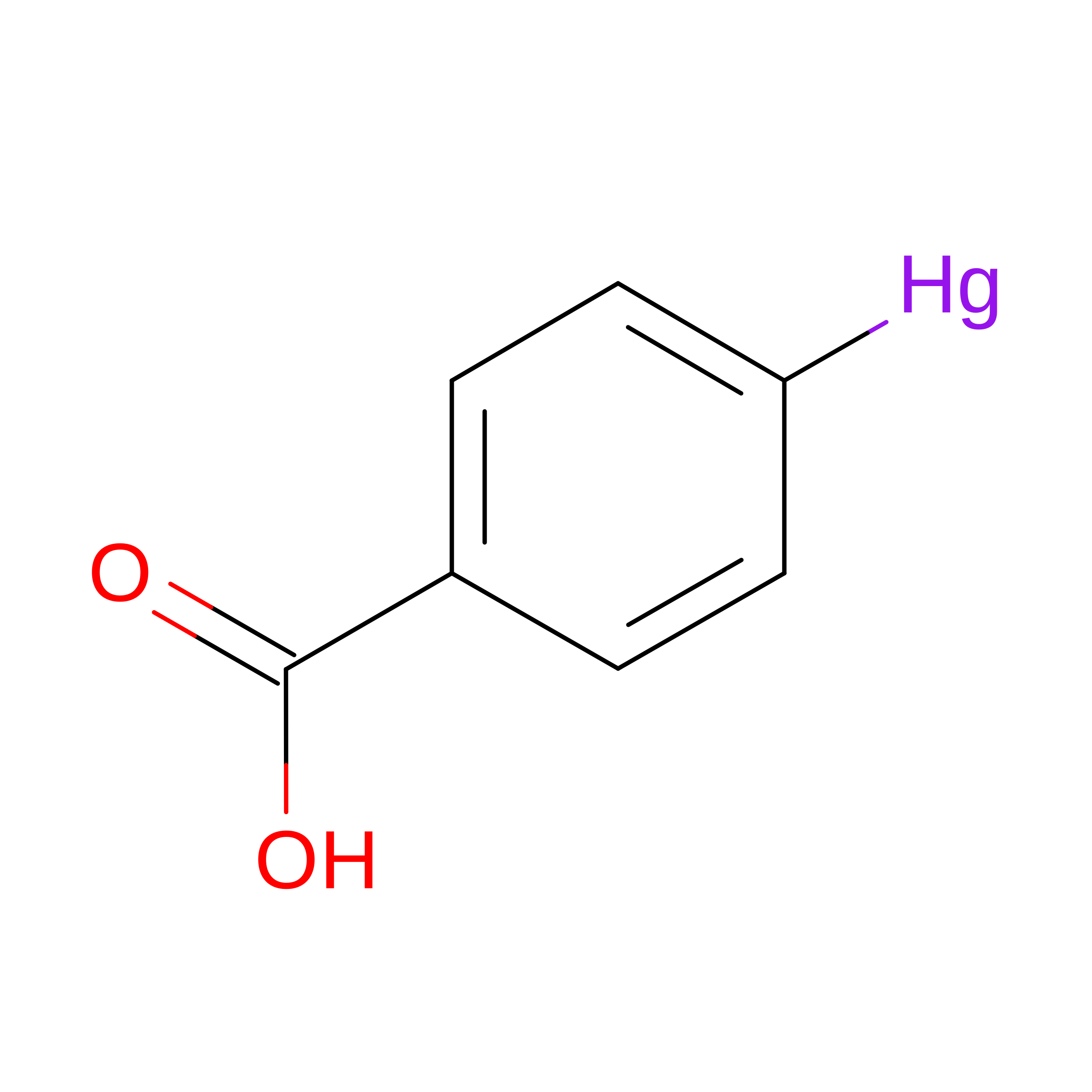
F [auth A]MERCURIBENZOIC ACID
C7 H5 Hg O2
FVFZSVRSDNUCGG-UHFFFAOYSA-N
ZN

Query on ZN


Download:Ideal Coordinates CCD File
B [auth A]ZINC ION
Zn
PTFCDOFLOPIGGS-UHFFFAOYSA-N
CL

Query on CL


Download:Ideal Coordinates CCD File
C [auth A]CHLORIDE ION
Cl
VEXZGXHMUGYJMC-UHFFFAOYSA-M

Experimental Data & Validation

Experimental Data

  • Method: X-RAY DIFFRACTION
  • Resolution: 1.46 Å
  • R-Value Free:  0.212 (Depositor), 0.208 (DCC) 
  • R-Value Work:  0.195 (Depositor), 0.190 (DCC) 
Space Group: P 1 21 1
Unit Cell:
Length ( Å )Angle ( ˚ )
a = 42.13α = 90
b = 41.42β = 104.5
c = 72.08γ = 90
Software Package:
Software NamePurpose
SCALEPACKdata scaling
CNSrefinement
CNSphasing

Structure Validation

View Full Validation Report


Ligand Structure Quality Assessment 


Entry History 

Deposition Data

Revision History  (Full details and data files)

  • Version 1.0: 2007-02-06
    Type: Initial release
  • Version 1.1: 2008-05-01
    Changes: Version format compliance
  • Version 1.2: 2011-07-13
    Changes: Version format compliance
  • Version 1.3: 2023-08-30
    Changes: Data collection, Database references, Derived calculations, Refinement description
  • Version 1.4: 2026-08-12
    Type: Remediation
    Reason: Metalloprotein remediation
    Changes: Derived calculations, Structure summary