2GD5

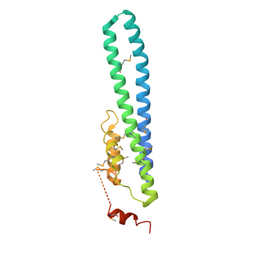
Structural basis for budding by the ESCRTIII factor CHMP3


Experimental Data Snapshot

  • Method: X-RAY DIFFRACTION
  • Resolution: 2.80 Å
  • R-Value Free: 0.301 
  • R-Value Work: 0.259 
  • R-Value Observed: 0.261 

wwPDB Validation   3D Report Full Report


This is version 1.3 of the entry. See complete history


Literature

Structural Basis for Budding by the ESCRT-III Factor CHMP3.

Pineda-Molina, E.Ravelli, R.B.Zamborlini, A.Usami, Y.Weissenhorn, W.

(2006) Dev Cell 10: 821-830

  • DOI: https://doi.org/10.1016/j.devcel.2006.03.013
  • Primary Citation of Related Structures:  
    2GD5

  • PubMed Abstract: 
  • The vacuolar protein sorting machinery regulates multivesicular body biogenesis and is selectively recruited by enveloped viruses to support budding. Here we report the crystal structure of the human ESCRT-III protein CHMP3 at 2.8 A resolution. The core structure of CHMP3 folds into a flat helical arrangement that assembles into a lattice, mainly via two different dimerization modes, and unilaterally exposes a highly basic surface ...

    The vacuolar protein sorting machinery regulates multivesicular body biogenesis and is selectively recruited by enveloped viruses to support budding. Here we report the crystal structure of the human ESCRT-III protein CHMP3 at 2.8 A resolution. The core structure of CHMP3 folds into a flat helical arrangement that assembles into a lattice, mainly via two different dimerization modes, and unilaterally exposes a highly basic surface. The C terminus, the target for Vps4-induced ESCRT disassembly, extends from the opposite side of the membrane targeting region. Mutations within the basic and dimerization regions hinder bilayer interaction in vivo and reverse the dominant-negative effect of a truncated CHMP3 fusion protein on HIV-1 budding. Thus, the final steps in the budding process may include CHMP protein polymerization and lattice formation on membranes by employing different bilayer-recognizing surfaces, a function shared by all CHMP family members.


    Organizational Affiliation

    European Molecular Biology Laboratory, 6 rue Jules Horowitz, 38042 Grenoble, France.


Macromolecules
Find similar proteins by:  (by identity cutoff)  |  3D Structure
Entity ID: 1
MoleculeChainsSequence LengthOrganismDetailsImage
Charged multivesicular body protein 3
A, B, C, D
179Homo sapiensMutation(s): 12 
Gene Names: CHMP3CGI149NEDFVPS24CGI-149
UniProt & NIH Common Fund Data Resources
Find proteins for Q9Y3E7 (Homo sapiens)
Explore Q9Y3E7 
Go to UniProtKB:  Q9Y3E7
PHAROS:  Q9Y3E7
Entity Groups  
Sequence Clusters30% Identity50% Identity70% Identity90% Identity95% Identity100% Identity
UniProt GroupQ9Y3E7
Protein Feature View
Expand
  • Reference Sequence
Small Molecules
Modified Residues  1 Unique
IDChainsTypeFormula2D DiagramParent
MSE
Query on MSE
A, B, C, D
L-PEPTIDE LINKINGC5 H11 N O2 SeMET
Experimental Data & Validation

Experimental Data

  • Method: X-RAY DIFFRACTION
  • Resolution: 2.80 Å
  • R-Value Free: 0.301 
  • R-Value Work: 0.259 
  • R-Value Observed: 0.261 
  • Space Group: P 1
Unit Cell:
Length ( Å )Angle ( ˚ )
a = 33.58α = 68.68
b = 72.57β = 83.61
c = 89.23γ = 76.7
Software Package:
Software NamePurpose
REFMACrefinement
ProDCdata collection
XDSdata scaling
SHELXSphasing

Structure Validation

View Full Validation Report


Entry History 

Deposition Data

Revision History  (Full details and data files)

  • Version 1.0: 2006-06-13
    Type: Initial release
  • Version 1.1: 2008-05-01
    Changes: Version format compliance
  • Version 1.2: 2011-07-13
    Changes: Advisory, Version format compliance
  • Version 1.3: 2017-10-18
    Changes: Refinement description