2FZU

Reduced enolate chromophore intermediate for GFP variant


Experimental Data Snapshot

  • Method: X-RAY DIFFRACTION
  • Resolution: 1.25 Å
  • R-Value Free: 0.199 
  • R-Value Work: 0.142 

wwPDB Validation   3D Report Full Report


This is version 1.5 of the entry. See complete history


Literature

Structural evidence for an enolate intermediate in GFP fluorophore biosynthesis.

Barondeau, D.P.Tainer, J.A.Getzoff, E.D.

(2006) J Am Chem Soc 128: 3166-3168

  • DOI: https://doi.org/10.1021/ja0552693
  • Primary Citation of Related Structures:  
    2FWQ, 2FZU

  • PubMed Abstract: 

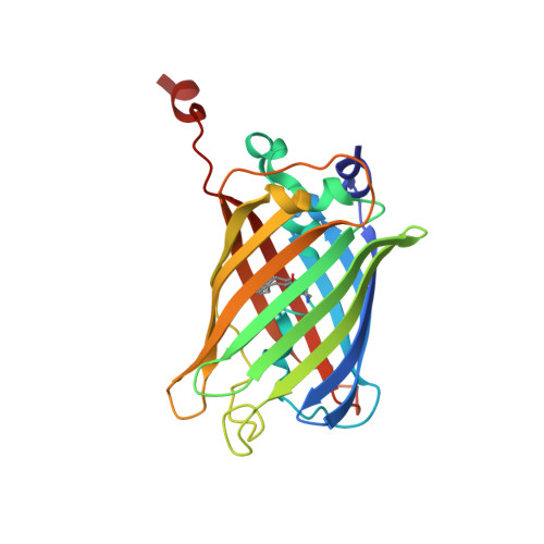
    The Aequorea victoria green fluorescent protein (GFP) creates a fluorophore from its component amino acids Ser65, Tyr66, and Gly67 through a remarkable post-translational modification, involving spontaneous peptide backbone cyclization, dehydration, and oxidation reactions. Here we test and extend the understanding of fluorophore biosynthesis by coupling chemical reduction and anaerobic methodologies with kinetic analyses and protein structure determination. Two high-resolution structures of dithionite-treated GFP variants reveal a previously uncharacterized enolate intermediate form of the chromophore that is viable in generating a fluorophore (t1/2 = 39 min-1) upon exposure to air. Isolation of this enolate intermediate will now allow specific probing of the rate-limiting oxidation step for fluorophore biosynthesis in GFP and its red fluorescent protein homologues. Such targeted characterizations may lead to the design of faster maturing proteins with enhanced applications in biotechnology and cell biology. Moreover, our results reveal how the GFP protein environment mimics enzyme systems, by stabilizing an otherwise high energy enolate intermediate to achieve its post-translational modification.


  • Organizational Affiliation

    Department of Molecular Biology, The Skaggs Institute for Chemical Biology, The Scripps Research Institute, 10550 North Torrey Pines Road, La Jolla, California 92037, USA.


Macromolecules
Find similar proteins by:  (by identity cutoff)  |  3D Structure
Entity ID: 1
MoleculeChains Sequence LengthOrganismDetailsImage
Green fluorescent protein237Aequorea victoriaMutation(s): 4 
Gene Names: GFP
UniProt
Find proteins for P42212 (Aequorea victoria)
Explore P42212 
Go to UniProtKB:  P42212
Entity Groups  
Sequence Clusters30% Identity50% Identity70% Identity90% Identity95% Identity100% Identity
UniProt GroupP42212
Sequence Annotations
Expand
  • Reference Sequence
Small Molecules
Modified Residues  1 Unique
IDChains TypeFormula2D DiagramParent
C12
Query on C12
A
L-PEPTIDE LINKINGC15 H18 N3 O4THR, TYR, GLY
Experimental Data & Validation

Experimental Data

  • Method: X-RAY DIFFRACTION
  • Resolution: 1.25 Å
  • R-Value Free: 0.199 
  • R-Value Work: 0.142 
  • Space Group: P 21 21 21
Unit Cell:
Length ( Å )Angle ( ˚ )
a = 51.35α = 90
b = 62.52β = 90
c = 71.75γ = 90
Software Package:
Software NamePurpose
DENZOdata reduction
SCALEPACKdata scaling
AMoREphasing
SHELXL-97refinement

Structure Validation

View Full Validation Report


Entry History 

Deposition Data

Revision History  (Full details and data files)

  • Version 1.0: 2006-03-14
    Type: Initial release
  • Version 1.1: 2008-05-01
    Changes: Version format compliance
  • Version 1.2: 2011-07-13
    Changes: Version format compliance
  • Version 1.3: 2021-10-20
    Changes: Database references, Derived calculations
  • Version 1.4: 2023-08-30
    Changes: Data collection, Refinement description
  • Version 1.5: 2023-11-15
    Changes: Data collection, Derived calculations