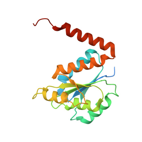
2FWP

Structure of PurE (N5-carboxyaminoimidazole ribonucleotide mutase) H59N from the acidophilic bacterium Acetobacter aceti, bound to isocair


Experimental Data Snapshot

  • Method: X-RAY DIFFRACTION
  • Resolution: 1.85 Å
  • R-Value Free: 0.225 
  • R-Value Work: 0.198 
  • R-Value Observed: 0.198 

Starting Model: experimental
View more details

wwPDB Validation   3D Report Full Report


Ligand Structure Quality Assessment 


This is version 1.4 of the entry. See complete history

Re-refinement Note

A newer entry is available that reflects an alternative modeling of the original data: 5CLJ


Literature

Biochemical and Structural Studies of N(5)-Carboxyaminoimidazole Ribonucleotide Mutase from the Acidophilic Bacterium Acetobacter aceti.

Constantine, C.Z.Starks, C.M.Mill, C.P.Ransome, A.E.Karpowicz, S.J.Francois, J.A.Goodman, R.A.Kappock, T.J.

(2006) Biochemistry 45: 8193-8208

  • DOI: https://doi.org/10.1021/bi060465n
  • Primary Citation of Related Structures:  
    2FW1, 2FW6, 2FW7, 2FW8, 2FW9, 2FWA, 2FWB, 2FWI, 2FWJ, 2FWP

  • PubMed Abstract: 

    N5-carboxyaminoimidazole ribonucleotide (N5-CAIR) mutase (PurE) catalyzes the reversible interconversion of acid-labile compounds N5-CAIR and 4-carboxy-5-aminoimidazole ribonucleotide (CAIR). We have examined PurE from the acidophilic bacterium Acetobacter aceti (AaPurE), focusing on its adaptation to acid pH and the roles of conserved residues His59 and His89. Both AaPurE and Escherichia coli PurE showed quasi-reversible acid-mediated inactivation, but wt AaPurE was much more stable at pH 3.5, with a > or = 20 degrees C higher thermal unfolding temperature at all pHs. His89 is not essential and does not function as part of a proton relay system. The kcat pH-rate profile was consistent with the assignment of pK1 to unproductive protonation of bound nucleotide and pK2 to deprotonation of His59. A 1.85 A resolution crystal structure of the inactive mutant H59N-AaPurE soaked in CAIR showed that protonation of CAIR C4 can occur in the absence of His59. The resulting species, modeled as isoCAIR [4(R)-carboxy-5-iminoimidazoline ribonucleotide], is strongly stabilized by extensive interactions with the enzyme and a water molecule. The carboxylate moiety is positioned in a small pocket proposed to facilitate nucleotide decarboxylation in the forward direction (N5-CAIR --> CAIR) [Meyer, E., Kappock, T. J., Osuji, C., and Stubbe, J. (1999) Biochemistry 38, 3012-3018]. Comparisons with model studies suggest that in the reverse (nonbiosynthetic) direction PurE favors protonation of CAIR C4. We suggest that the essential role of protonated His59 is to lower the barrier to decarboxylation by stabilizing a CO2-azaenolate intermediate.


  • Organizational Affiliation

    Department of Chemistry, Washington University, St. Louis, Missouri 63130-4899, USA.


Macromolecules
Find similar proteins by:  (by identity cutoff)  |  3D Structure
Entity ID: 1
MoleculeChains Sequence LengthOrganismDetailsImage
N5-carboxyaminoimidazole ribonucleotide mutase
A, B
183Acetobacter acetiMutation(s): 1 
Gene Names: purE
UniProt
Find proteins for Q2QJL3 (Acetobacter aceti)
Explore Q2QJL3 
Go to UniProtKB:  Q2QJL3
Entity Groups  
Sequence Clusters30% Identity50% Identity70% Identity90% Identity95% Identity100% Identity
UniProt GroupQ2QJL3
Sequence Annotations
Expand
  • Reference Sequence
Small Molecules
Ligands 2 Unique
IDChains Name / Formula / InChI Key2D Diagram3D Interactions
ICR
Query on ICR

Download Ideal Coordinates CCD File 
C [auth A](4R)-5-IMINO-1-(5-O-PHOSPHONO-BETA-D-RIBOFURANOSYL)-4,5-DIHYDRO-1H-IMIDAZOLE-4-CARBOXYLIC ACID
C9 H14 N3 O9 P
WYRPDIVLQIXBHB-IUONDULESA-N
CIT
Query on CIT

Download Ideal Coordinates CCD File 
D [auth B]CITRIC ACID
C6 H8 O7
KRKNYBCHXYNGOX-UHFFFAOYSA-N
Binding Affinity Annotations 
IDSourceBinding Affinity
CIT PDBBind:  2FWP Kd: 1.20e+6 (nM) from 1 assay(s)
Experimental Data & Validation

Experimental Data

Unit Cell:
Length ( Å )Angle ( ˚ )
a = 98.683α = 90
b = 98.683β = 90
c = 165.59γ = 90
Software Package:
Software NamePurpose
CNSrefinement
HKL-2000data reduction
SCALEPACKdata scaling
CNSphasing

Structure Validation

View Full Validation Report


Ligand Structure Quality Assessment 


Entry History 

Deposition Data

Revision History  (Full details and data files)

  • Version 1.0: 2006-06-13
    Type: Initial release
  • Version 1.1: 2008-05-01
    Changes: Version format compliance
  • Version 1.2: 2011-07-13
    Changes: Version format compliance
  • Version 1.3: 2021-10-20
    Changes: Database references, Derived calculations
  • Version 1.4: 2023-08-30
    Changes: Data collection, Refinement description