2FG4

Structure of Human Ferritin L Chain


Experimental Data Snapshot

  • Method: X-RAY DIFFRACTION
  • Resolution: 2.10 Å
  • R-Value Free: 0.220 
  • R-Value Work: 0.194 

wwPDB Validation   3D Report Full Report


This is version 1.3 of the entry. See complete history


Literature

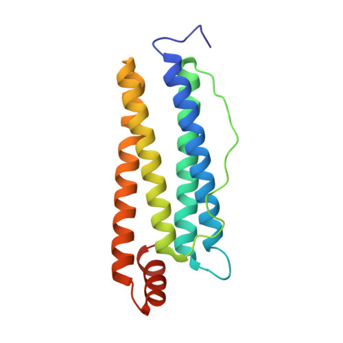
Structure of human ferritin L chain.

Wang, Z.Li, C.Ellenburg, M.Soistman, E.Ruble, J.Wright, B.Ho, J.X.Carter, D.C.

(2006) Acta Crystallogr D Biol Crystallogr 62: 800-806

  • DOI: https://doi.org/10.1107/S0907444906018294
  • Primary Citation of Related Structures:  
    2FFX, 2FG4, 2FG8

  • PubMed Abstract: 
  • Ferritin is the major iron-storage protein present in all cells. It generally contains 24 subunits, with different ratios of heavy chain (H) to light chain (L), in the shape of a hollow sphere hosting up to 4500 ferric Fe atoms inside. H-rich ferritins catalyse the oxidation of iron(II), while L-rich ferritins promote the nucleation and storage of iron(III) ...

    Ferritin is the major iron-storage protein present in all cells. It generally contains 24 subunits, with different ratios of heavy chain (H) to light chain (L), in the shape of a hollow sphere hosting up to 4500 ferric Fe atoms inside. H-rich ferritins catalyse the oxidation of iron(II), while L-rich ferritins promote the nucleation and storage of iron(III). Several X-ray structures have been determined, including those of L-chain ferritins from horse spleen (HoSF), recombinant L-chain ferritins from horse (HoLF), mouse (MoLF) and bullfrog (BfLF) as well as recombinant human H-chain ferritin (HuHF). Here, structures have been determined of two crystal forms of recombinant human L-chain ferritin (HuLF) obtained from native and perdeuterated proteins. The structures show a cluster of acidic residues at the ferrihydrite nucleation site and at the iron channel along the threefold axis. An ordered Cd2+ structure is observed within the iron channel, offering further insight into the route and mechanism of iron transport into the capsid. The loop between helices D and E, which is disordered in many other L-chain structures, is clearly visible in these two structures. The crystals generated from perdeuterated HuLF will be used for neutron diffraction studies.


    Organizational Affiliation

    New Century Pharmaceuticals Inc., 895 Martin Road, Huntsville, Alabama 35824, USA.


Macromolecules
Find similar proteins by:  (by identity cutoff)  |  3D Structure
Entity ID: 1
MoleculeChainsSequence LengthOrganismDetailsImage
Ferritin light chainA174Homo sapiensMutation(s): 0 
Gene Names: FTL
UniProt & NIH Common Fund Data Resources
Find proteins for P02792 (Homo sapiens)
Explore P02792 
Go to UniProtKB:  P02792
PHAROS:  P02792
Entity Groups  
Sequence Clusters30% Identity50% Identity70% Identity90% Identity95% Identity100% Identity
UniProt GroupP02792
Protein Feature View
Expand
  • Reference Sequence
Experimental Data & Validation

Experimental Data

  • Method: X-RAY DIFFRACTION
  • Resolution: 2.10 Å
  • R-Value Free: 0.220 
  • R-Value Work: 0.194 
  • Space Group: I 4 3 2
Unit Cell:
Length ( Å )Angle ( ˚ )
a = 152.865α = 90
b = 152.865β = 90
c = 152.865γ = 90
Software Package:
Software NamePurpose
CNXrefinement
DENZOdata reduction
SCALEPACKdata scaling
AMoREphasing

Structure Validation

View Full Validation Report


Entry History 

Deposition Data

Revision History  (Full details and data files)

  • Version 1.0: 2006-07-04
    Type: Initial release
  • Version 1.1: 2008-05-01
    Changes: Version format compliance
  • Version 1.2: 2011-07-13
    Changes: Derived calculations, Version format compliance
  • Version 1.3: 2011-11-16
    Changes: Atomic model