2FBD | pdb_00002fbd

The crystallographic structure of the digestive lysozyme 1 from Musca domestica at 1.90 Ang.


Experimental Data Snapshot

  • Method: X-RAY DIFFRACTION
  • Resolution: 1.90 Å
  • R-Value Free: 
    0.198 (Depositor), 0.159 (DCC) 
  • R-Value Work: 
    0.153 (Depositor), 0.158 (DCC) 
  • R-Value Observed: 
    0.156 (Depositor) 

Starting Model: experimental
View more details

wwPDB Validation 3D Report Full Report

Validation slider image for 2FBD

This is version 1.5 of the entry. See complete history

Literature

Crystallization, data collection and phasing of two digestive lysozymes from Musca domestica.

Marana, S.R.Cancado, F.C.Valero, A.A.Ferreira, C.Terra, W.R.Barbosa, J.A.R.G.

(2006) Acta Crystallogr Sect F Struct Biol Cryst Commun 62: 750-752

  • DOI: https://doi.org/10.1107/S1744309106024201
  • Primary Citation Related Structures: 
    2FBD, 2H5Z

  • PubMed Abstract: 

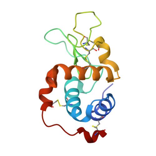
    Lysozymes are mostly known for their defensive role against bacteria, but in several animals lysozymes have a digestive function. Here, the initial crystallographic characterization of two digestive lysozymes from Musca domestica are presented. The proteins were crystallized using the sitting-drop vapour-diffusion method in the presence of ammonium sulfate or PEG/2-propanol as the precipitant. X-ray diffraction data were collected to a maximum resolution of 1.9 angstroms using synchrotron radiation. The lysozyme 1 and 2 crystals belong to the monoclinic space group P2(1) (unit-cell parameters a = 36.52, b = 79.44, c = 45.20 angstroms, beta = 102.97 degrees) and the orthorhombic space group P2(1)2(1)2 (unit-cell parameters a = 73.90, b = 96.40, c = 33.27 angstroms), respectively. The crystal structures were solved by molecular replacement and structure refinement is in progress.


  • Organizational Affiliation
    • Departamento de Bioquímica, Instituto de Química, Universidade de São Paulo, São Paulo, Brazil.

Macromolecule Content 

  • Total Structure Weight: 28.16 kDa 
  • Atom Count: 2,316 
  • Modeled Residue Count: 244 
  • Deposited Residue Count: 244 
  • Unique protein chains: 1

Macromolecules

Find similar proteins by:|  3D Structure
Entity ID: 1
MoleculeChains  Sequence LengthOrganismDetailsImage
Lysozyme 1
A, B
122Musca domesticaMutation(s): 0 
Gene Names: AY344589
EC: 3.2.1.17
UniProt
Find proteins for Q7YT16 (Musca domestica)
Explore Q7YT16 
Go to UniProtKB:  Q7YT16
Entity Groups
Sequence Clusters30% Identity50% Identity70% Identity90% Identity95% Identity100% Identity
UniProt GroupQ7YT16
Sequence Annotations
Expand
Reference Sequence

Experimental Data & Validation

Experimental Data

  • Method: X-RAY DIFFRACTION
  • Resolution: 1.90 Å
  • R-Value Free:  0.198 (Depositor), 0.159 (DCC) 
  • R-Value Work:  0.153 (Depositor), 0.158 (DCC) 
  • R-Value Observed: 0.156 (Depositor) 
Space Group: P 1 21 1
Unit Cell:
Length ( Å )Angle ( ˚ )
a = 36.523α = 90
b = 79.435β = 102.97
c = 45.203γ = 90
Software Package:
Software NamePurpose
REFMACrefinement
HKL-2000data reduction
HKL-2000data scaling
MOLREPphasing

Structure Validation

View Full Validation Report


Entry History 

Deposition Data

Revision History  (Full details and data files)

  • Version 1.0: 2006-12-12
    Type: Initial release
  • Version 1.1: 2008-05-01
    Changes: Version format compliance
  • Version 1.2: 2011-07-13
    Changes: Advisory, Derived calculations, Version format compliance
  • Version 1.3: 2017-06-21
    Changes: Database references
  • Version 1.4: 2023-08-30
    Changes: Data collection, Database references, Derived calculations, Refinement description
  • Version 1.5: 2024-10-30
    Changes: Structure summary