2EWW

Crystal Structure of the Pilus Retraction Motor PilT and Bound ATP


Experimental Data Snapshot

  • Method: X-RAY DIFFRACTION
  • Resolution: 3.20 Å
  • R-Value Free: 0.310 
  • R-Value Work: 0.239 
  • R-Value Observed: 0.242 

wwPDB Validation   3D Report Full Report


Ligand Structure Quality Assessment 


This is version 1.2 of the entry. See complete history


Literature

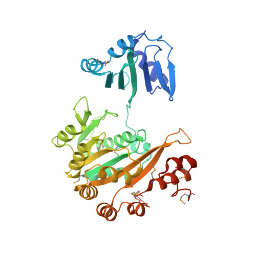
Crystal structures of the pilus retraction motor PilT suggest large domain movements and subunit cooperation drive motility.

Satyshur, K.A.Worzalla, G.A.Meyer, L.S.Heiniger, E.K.Aukema, K.G.Misic, A.M.Forest, K.T.

(2007) Structure 15: 363-376

  • DOI: https://doi.org/10.1016/j.str.2007.01.018
  • Primary Citation of Related Structures:  
    2EWV, 2EWW, 2EYU, 2GSZ

  • PubMed Abstract: 

    PilT is a hexameric ATPase required for bacterial type IV pilus retraction and surface motility. Crystal structures of ADP- and ATP-bound Aquifex aeolicus PilT at 2.8 and 3.2 A resolution show N-terminal PAS-like and C-terminal RecA-like ATPase domains followed by a set of short C-terminal helices. The hexamer is formed by extensive polar subunit interactions between the ATPase core of one monomer and the N-terminal domain of the next. An additional structure captures a nonsymmetric PilT hexamer in which approach of invariant arginines from two subunits to the bound nucleotide forms an enzymatically competent active site. A panel of pilT mutations highlights the importance of the arginines, the PAS-like domain, the polar subunit interface, and the C-terminal helices for retraction. We present a model for ATP binding leading to dramatic PilT domain motions, engagement of the arginine wire, and subunit communication in this hexameric motor. Our conclusions apply to the entire type II/IV secretion ATPase family.


  • Organizational Affiliation

    Department of Bacteriology, University of Wisconsin-Madison, Madison, WI 53706, USA.


Macromolecules
Find similar proteins by:  (by identity cutoff)  |  3D Structure
Entity ID: 1
MoleculeChains Sequence LengthOrganismDetailsImage
twitching motility protein PilT372Aquifex aeolicusMutation(s): 10 
UniProt
Find proteins for O66950 (Aquifex aeolicus (strain VF5))
Explore O66950 
Go to UniProtKB:  O66950
Entity Groups  
Sequence Clusters30% Identity50% Identity70% Identity90% Identity95% Identity100% Identity
UniProt GroupO66950
Sequence Annotations
Expand
  • Reference Sequence
Small Molecules
Ligands 1 Unique
IDChains Name / Formula / InChI Key2D Diagram3D Interactions
ATP
Query on ATP

Download Ideal Coordinates CCD File 
B [auth A]ADENOSINE-5'-TRIPHOSPHATE
C10 H16 N5 O13 P3
ZKHQWZAMYRWXGA-KQYNXXCUSA-N
Modified Residues  1 Unique
IDChains TypeFormula2D DiagramParent
MSE
Query on MSE
A
L-PEPTIDE LINKINGC5 H11 N O2 SeMET
Experimental Data & Validation

Experimental Data

  • Method: X-RAY DIFFRACTION
  • Resolution: 3.20 Å
  • R-Value Free: 0.310 
  • R-Value Work: 0.239 
  • R-Value Observed: 0.242 
  • Space Group: P 6
Unit Cell:
Length ( Å )Angle ( ˚ )
a = 107.38α = 90
b = 107.38β = 90
c = 68.86γ = 120
Software Package:
Software NamePurpose
REFMACrefinement
MOSFLMdata reduction
CCP4data scaling
SHARPphasing

Structure Validation

View Full Validation Report


Ligand Structure Quality Assessment 


Entry History 

Deposition Data

Revision History  (Full details and data files)

  • Version 1.0: 2006-11-21
    Type: Initial release
  • Version 1.1: 2008-05-01
    Changes: Version format compliance
  • Version 1.2: 2011-07-13
    Changes: Version format compliance