2EU2 | pdb_00002eu2

Human Carbonic Anhydrase II in complex with novel inhibitors


Experimental Data Snapshot

  • Method: X-RAY DIFFRACTION
  • Resolution: 1.15 Å
  • R-Value Free: 
    0.166 (Depositor), 0.138 (DCC) 
  • R-Value Work: 
    0.136 (Depositor), 0.143 (DCC) 
  • R-Value Observed: 
    0.136 (Depositor) 

Starting Model: experimental
View more details

wwPDB Validation 3D Report Full Report

Validation slider image for 2EU2

Ligand Structure Quality Assessment 


This is version 1.6 of the entry. See complete history

Literature

X-ray crystallographic studies reveal that the incorporation of spacer groups in carbonic anhydrase inhibitors causes alternate binding modes.

Fisher, S.Z.Govindasamy, L.Boyle, N.Agbandje-McKenna, M.Silverman, D.N.Blackburn, G.M.McKenna, R.

(2006) Acta Crystallogr Sect F Struct Biol Cryst Commun 62: 618-622

  • DOI: https://doi.org/10.1107/S1744309106020446
  • Primary Citation Related Structures: 
    2EU2, 2EU3

  • PubMed Abstract: 

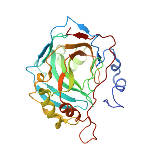
    Human carbonic anhydrases (CAs) are well studied targets for the development of inhibitors for pharmaceutical applications. The crystal structure of human CA II has been determined in complex with two CA inhibitors (CAIs) containing conventional sulfonamide and thiadiazole moieties separated by a -CF2- or -CHNH2- spacer group. The structures presented here reveal that these spacer groups allow novel binding modes for the thiadiazole moiety compared with conventional CAIs.


  • Organizational Affiliation
    • Department of Biochemistry and Molecular Biology, College of Medicine, University of Florida, Gainesville, FL 32610, USA.

Macromolecule Content 

  • Total Structure Weight: 29.59 kDa 
  • Atom Count: 2,384 
  • Modeled Residue Count: 257 
  • Deposited Residue Count: 260 
  • Unique protein chains: 1

Macromolecules

Find similar proteins by:|  3D Structure
Entity ID: 1
MoleculeChains  Sequence LengthOrganismDetailsImage
Carbonic anhydrase 2260Homo sapiensMutation(s): 0 
Gene Names: CA2
EC: 4.2.1.1 (PDB Primary Data), 4.2.1.69 (UniProt)
UniProt & NIH Common Fund Data Resources
Find proteins for P00918 (Homo sapiens)
Explore P00918 
Go to UniProtKB:  P00918
PHAROS:  P00918
GTEx:  ENSG00000104267 
Entity Groups
Sequence Clusters30% Identity50% Identity70% Identity90% Identity95% Identity100% Identity
UniProt GroupP00918
Sequence Annotations
Expand
Reference Sequence

Small Molecules

Ligands 2 Unique
IDChains Name / Formula / InChI Key2D Diagram3D Interactions
5DS

Query on 5DS


Download:Ideal Coordinates CCD File
C [auth A](R)-1-AMINO-1-[5-(DIMETHYLAMINO)-1,3,4-THIADIAZOL-2-YL]METHANESULFONAMIDE
C5 H11 N5 O2 S2
PBUMATDKSRINDG-GSVOUGTGSA-N
ZN

Query on ZN


Download:Ideal Coordinates CCD File
B [auth A]ZINC ION
Zn
PTFCDOFLOPIGGS-UHFFFAOYSA-N

Experimental Data & Validation

Experimental Data

  • Method: X-RAY DIFFRACTION
  • Resolution: 1.15 Å
  • R-Value Free:  0.166 (Depositor), 0.138 (DCC) 
  • R-Value Work:  0.136 (Depositor), 0.143 (DCC) 
  • R-Value Observed: 0.136 (Depositor) 
Space Group: P 1 21 1
Unit Cell:
Length ( Å )Angle ( ˚ )
a = 42.965α = 90
b = 42.057β = 104.03
c = 72.412γ = 90
Software Package:
Software NamePurpose
SHELXL-97refinement
SCALEPACKdata scaling
CNSrefinement
HKL-2000data reduction
CNSphasing
HKL-2000data collection

Structure Validation

View Full Validation Report


Ligand Structure Quality Assessment 


Entry History 

Deposition Data

Revision History  (Full details and data files)

  • Version 1.0: 2006-07-11
    Type: Initial release
  • Version 1.1: 2008-05-01
    Changes: Version format compliance
  • Version 1.2: 2011-07-13
    Changes: Version format compliance
  • Version 1.3: 2017-10-18
    Changes: Refinement description
  • Version 1.4: 2019-07-24
    Changes: Data collection, Refinement description
  • Version 1.5: 2019-08-14
    Changes: Data collection
  • Version 1.6: 2023-08-23
    Changes: Data collection, Database references, Derived calculations, Refinement description