2DPQ

The crystal structures of the calcium-bound con-G and con-T(K7gamma) dimeric peptides demonstrate a novel metal-dependent helix-forming motif


Experimental Data Snapshot

  • Method: X-RAY DIFFRACTION
  • Resolution: 1.25 Å
  • R-Value Free: 0.161 
  • R-Value Work: 0.119 
  • R-Value Observed: 0.119 

wwPDB Validation   3D Report Full Report


This is version 1.5 of the entry. See complete history


Literature

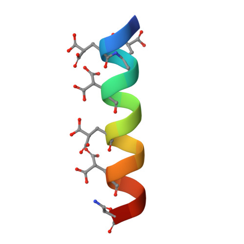
The crystal structures of the calcium-bound con-G and con-T[K7gamma] dimeric peptides demonstrate a metal-dependent helix-forming motif

Cnudde, S.E.Prorok, M.Dai, Q.Castellino, F.J.Geiger, J.H.

(2007) J Am Chem Soc 129: 1586-1593

  • DOI: https://doi.org/10.1021/ja065722q
  • Primary Citation of Related Structures:  
    2DPQ, 2DPR

  • PubMed Abstract: 

    Short peptides that have the ability to form stable alpha-helices in solution are rare, and a number of strategies have been used to produce them, including the use of metal chelation to stabilize folding of the backbone. However, no example exists of a structurally well-defined helix stabilized exclusively through metal ion chelation. Conantokins (con)-G and -T are short peptides that are potent antagonists of N-methyl-D-aspartate receptor channels. While con-G exhibits no helicity alone, it undergoes a structural transition to a helical conformation in the presence of a variety of multivalent cations, especially Mg2+ and Ca2+. This complexation also results in antiparallel dimerization of two peptide helices in the presence of Ca2+, but not Mg2+. A con-T variant, con-T[K7gamma], displays very similar behavior. We have solved the crystal structures of both Ca2+/con-G and Ca2+/con-T [K7gamma] at atomic resolution. These structures clearly show the nature of the metal-dependent dimerization and helix formation and surprisingly also show that the con-G dimer interface is completely different from the con-T[K7gamma] interface, even though the metal chelation is similar in the two peptides. This represents a new paradigm in helix stabilization completely independent of the hydrophobic effect, which we define as the "metallo-zipper."


  • Organizational Affiliation

    Department of Biochemistry, Michigan State University, East Lansing, Michigan 48824, USA.


Macromolecules

Find similar proteins by:  Sequence   |   3D Structure  

Entity ID: 1
MoleculeChains Sequence LengthOrganismDetailsImage
Conantokin-G18Conus geographusMutation(s): 1 
UniProt
Find proteins for P07231 (Conus geographus)
Explore P07231 
Go to UniProtKB:  P07231
Entity Groups  
Sequence Clusters30% Identity50% Identity70% Identity90% Identity95% Identity100% Identity
UniProt GroupP07231
Sequence Annotations
Expand
  • Reference Sequence
Experimental Data & Validation

Experimental Data

  • Method: X-RAY DIFFRACTION
  • Resolution: 1.25 Å
  • R-Value Free: 0.161 
  • R-Value Work: 0.119 
  • R-Value Observed: 0.119 
  • Space Group: P 42 2 2
Unit Cell:
Length ( Å )Angle ( ˚ )
a = 29.334α = 90
b = 29.334β = 90
c = 46.892γ = 90
Software Package:
Software NamePurpose
DENZOdata reduction
SCALEPACKdata scaling
SHELXrefinement
PDB_EXTRACTdata extraction
PHASERphasing
SHELXLrefinement

Structure Validation

View Full Validation Report


Entry History 

Deposition Data

Revision History  (Full details and data files)

  • Version 1.0: 2007-04-24
    Type: Initial release
  • Version 1.1: 2008-04-01
    Changes: Version format compliance
  • Version 1.2: 2011-07-13
    Changes: Derived calculations, Version format compliance
  • Version 1.3: 2013-07-24
    Changes: Database references
  • Version 1.4: 2017-10-11
    Changes: Refinement description
  • Version 1.5: 2024-04-03
    Changes: Data collection, Database references, Derived calculations, Refinement description