2D1J

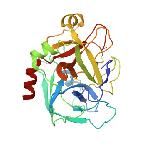
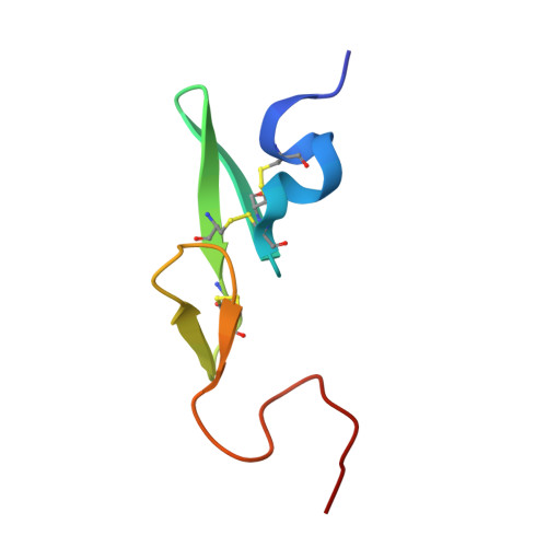
Factor Xa in complex with the inhibitor 2-[[4-[(5-chloroindol-2-yl)sulfonyl]piperazin-1-yl] carbonyl]thieno[3,2-b]pyridine n-oxide


Experimental Data Snapshot

  • Method: X-RAY DIFFRACTION
  • Resolution: 2.20 Å
  • R-Value Free: 0.232 
  • R-Value Work: 0.186 
  • R-Value Observed: 0.188 

Starting Model: experimental
View more details

wwPDB Validation   3D Report Full Report


This is version 1.3 of the entry. See complete history


Literature

Design, synthesis, and biological activity of non-basic compounds as factor Xa inhibitors: SAR study of S1 and aryl binding sites.

Komoriya, S.Haginoya, N.Kobayashi, S.Nagata, T.Mochizuki, A.Suzuki, M.Yoshino, T.Horino, H.Nagahara, T.Suzuki, M.Isobe, Y.Furugoori, T.

(2005) Bioorg Med Chem 13: 3927-3954

  • DOI: https://doi.org/10.1016/j.bmc.2005.04.006
  • Primary Citation of Related Structures:  
    1WU1, 2D1J

  • PubMed Abstract: 

    Compound 7 was identified as the active metabolite of 6 by HPLC and mass spectral analysis. Modification of lead compound 7 by transformation of its N-oxide 6-6 biaryl ring system and fused aromatics produced a series of non-basic fXa inhibitors with excellent potency in anti-fXa and anticoagulant assays. The optimized compounds 73b and 75b showed sub to one digit micromolar anticoagulant activity (PTCT2). Particularly, anti-fXa activity was detected in plasma of rats orally administered with 1mg/kg of compound 75b.


  • Organizational Affiliation

    Tokyo R&D Center, Daiichi Pharmaceutical Co. Ltd, 16-13 Kita-Kasai 1-Chome, Edogawa-ku, Tokyo 134-8630, Japan. komor0ni@daiichipharm.co.jp


Macromolecules
Find similar proteins by:  (by identity cutoff)  |  3D Structure
Entity ID: 1
MoleculeChains Sequence LengthOrganismDetailsImage
Coagulation factor X, heavy chain233Homo sapiensMutation(s): 0 
EC: 3.4.21.6
UniProt & NIH Common Fund Data Resources
Find proteins for P00742 (Homo sapiens)
Explore P00742 
Go to UniProtKB:  P00742
PHAROS:  P00742
GTEx:  ENSG00000126218 
Entity Groups  
Sequence Clusters30% Identity50% Identity70% Identity90% Identity95% Identity100% Identity
UniProt GroupP00742
Sequence Annotations
Expand
  • Reference Sequence
Find similar proteins by:  (by identity cutoff)  |  3D Structure
Entity ID: 2
MoleculeChains Sequence LengthOrganismDetailsImage
Coagulation factor X, light chain54Homo sapiensMutation(s): 0 
EC: 3.4.21.6
UniProt & NIH Common Fund Data Resources
Find proteins for P00742 (Homo sapiens)
Explore P00742 
Go to UniProtKB:  P00742
PHAROS:  P00742
GTEx:  ENSG00000126218 
Entity Groups  
Sequence Clusters30% Identity50% Identity70% Identity90% Identity95% Identity100% Identity
UniProt GroupP00742
Sequence Annotations
Expand
  • Reference Sequence
Small Molecules
Ligands 2 Unique
IDChains Name / Formula / InChI Key2D Diagram3D Interactions
D01
Query on D01

Download Ideal Coordinates CCD File 
E [auth A]2-({4-[(5-CHLORO-1H-INDOL-2-YL)SULFONYL]PIPERAZIN-1-YL}CARBONYL)THIENO[3,2-B]PYRIDINE 4-OXIDE
C20 H17 Cl N4 O4 S2
DHDQMXPAANQKDC-UHFFFAOYSA-N
CA
Query on CA

Download Ideal Coordinates CCD File 
C [auth A],
D [auth A]
CALCIUM ION
Ca
BHPQYMZQTOCNFJ-UHFFFAOYSA-N
Experimental Data & Validation

Experimental Data

  • Method: X-RAY DIFFRACTION
  • Resolution: 2.20 Å
  • R-Value Free: 0.232 
  • R-Value Work: 0.186 
  • R-Value Observed: 0.188 
  • Space Group: P 21 21 21
Unit Cell:
Length ( Å )Angle ( ˚ )
a = 57.078α = 90
b = 73.122β = 90
c = 79.949γ = 90
Software Package:
Software NamePurpose
d*TREKdata scaling
d*TREKdata reduction
REFMACrefinement

Structure Validation

View Full Validation Report


Entry History 

Deposition Data

  • Released Date: 2006-08-24 
  • Deposition Author(s): Suzuki, M.

Revision History  (Full details and data files)

  • Version 1.0: 2006-08-24
    Type: Initial release
  • Version 1.1: 2008-04-30
    Changes: Version format compliance
  • Version 1.2: 2011-07-13
    Changes: Version format compliance
  • Version 1.3: 2023-10-25
    Changes: Data collection, Database references, Derived calculations, Refinement description