2CN6 | pdb_00002cn6

Recombinant human H ferritin, K86Q and E107D mutant, soaked with Zn ions


Experimental Data Snapshot

  • Method: X-RAY DIFFRACTION
  • Resolution: 2.20 Å
  • R-Value Free: 
    0.230 (Depositor), 0.229 (DCC) 
  • R-Value Work: 
    0.195 (Depositor), 0.192 (DCC) 
  • R-Value Observed: 
    0.196 (Depositor) 

Starting Model: experimental
View more details

wwPDB Validation 3D Report Full Report

Validation slider image for 2CN6

This is version 1.4 of the entry. See complete history

Literature

High-Resolution X-Ray Structures of Human Apoferritin H-Chain Mutants Correlated with Their Activity and Metal-Binding Sites.

Toussaint, L.Bertrand, L.Hue, L.Crichton, R.R.Declercq, J.P.

(2007) J Mol Biology 365: 440

  • DOI: https://doi.org/10.1016/j.jmb.2006.10.010
  • Primary Citation Related Structures: 
    2CEI, 2CHI, 2CIH, 2CLU, 2CN6, 2CN7, 2IU2

  • PubMed Abstract: 

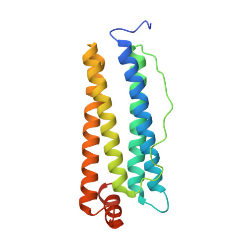
    Ferritins are a family of proteins distributed widely in nature. In bacterial, plant, and animal cells, ferritin appears to serve as a soluble, bioavailable, and non-toxic form of iron provider. Ferritins from animal sources are heteropolymers composed of two types of subunit, H and L, which differ mainly by the presence (H) or absence (L) of active ferroxidase centres. We report the crystallographic structures of four human H apoferritin variants at a resolution of up to 1.5 Angstrom. Crystal derivatives using Zn(II) as redox-stable alternative for Fe(II), allows us to characterize the different metal-binding sites. The ferroxidase centre, which is composed of sites A and B, binds metal with a preference for the A site. In addition, distinct Zn(II)-binding sites were found in the 3-fold axes, 4-fold axes and on the cavity surface near the ferroxidase centre. To study the importance of the distance of the two metal atoms in the ferroxidase centre, single and double replacement of glutamate 27 (site A) and glutamate 107 (site B), the two axial ligands, by aspartate residues have been carried out. The consequences for metal binding and the correlation with Fe(II) oxidation rates are discussed.


  • Organizational Affiliation
    • Biochemistry Unit, Université Catholique de Louvain, 1 Place Louis Pasteur, 1348 Louvain la Neuve, Belgium.

Macromolecule Content 

  • Total Structure Weight: 21.83 kDa 
  • Atom Count: 1,495 
  • Modeled Residue Count: 172 
  • Deposited Residue Count: 183 
  • Unique protein chains: 1

Macromolecules

Find similar proteins by:|  3D Structure
Entity ID: 1
MoleculeChains  Sequence LengthOrganismDetailsImage
FERRITIN HEAVY CHAIN183Homo sapiensMutation(s): 2 
EC: 1.16.3.1
UniProt & NIH Common Fund Data Resources
Find proteins for P02794 (Homo sapiens)
Explore P02794 
Go to UniProtKB:  P02794
PHAROS:  P02794
GTEx:  ENSG00000167996 
Entity Groups
Sequence Clusters30% Identity50% Identity70% Identity90% Identity95% Identity100% Identity
UniProt GroupP02794
Sequence Annotations
Expand
Reference Sequence

Small Molecules

Ligands 3 Unique
IDChains Name / Formula / InChI Key2D Diagram3D Interactions
GOL

Query on GOL


Download:Ideal Coordinates CCD File
J [auth A]GLYCEROL
C3 H8 O3
PEDCQBHIVMGVHV-UHFFFAOYSA-N
ZN

Query on ZN


Download:Ideal Coordinates CCD File
B [auth A]
D [auth A]
E [auth A]
F [auth A]
G [auth A]
B [auth A],
D [auth A],
E [auth A],
F [auth A],
G [auth A],
H [auth A],
I [auth A]
ZINC ION
Zn
PTFCDOFLOPIGGS-UHFFFAOYSA-N
CA

Query on CA


Download:Ideal Coordinates CCD File
C [auth A]CALCIUM ION
Ca
BHPQYMZQTOCNFJ-UHFFFAOYSA-N

Experimental Data & Validation

Experimental Data

  • Method: X-RAY DIFFRACTION
  • Resolution: 2.20 Å
  • R-Value Free:  0.230 (Depositor), 0.229 (DCC) 
  • R-Value Work:  0.195 (Depositor), 0.192 (DCC) 
  • R-Value Observed: 0.196 (Depositor) 
Space Group: F 4 3 2
Unit Cell:
Length ( Å )Angle ( ˚ )
a = 178.453α = 90
b = 178.453β = 90
c = 178.453γ = 90
Software Package:
Software NamePurpose
REFMACrefinement
XDSdata reduction
XSCALEdata scaling

Structure Validation

View Full Validation Report


Entry History 

Deposition Data

Revision History  (Full details and data files)

  • Version 1.0: 2006-12-13
    Type: Initial release
  • Version 1.1: 2011-05-08
    Changes: Version format compliance
  • Version 1.2: 2011-07-13
    Changes: Version format compliance
  • Version 1.3: 2019-05-08
    Changes: Data collection, Experimental preparation, Other
  • Version 1.4: 2023-12-13
    Changes: Data collection, Database references, Derived calculations, Other, Refinement description