


Structure of Lysozyme
Kleywegt, G.J., Divne, C.To be published.
Experimental Data Snapshot
Starting Model: experimental
View more details
wwPDB Validation 3D Report Full Report

Entity ID: 1 | |||||
|---|---|---|---|---|---|
| Molecule | Chains | Sequence Length | Organism | Details | Image |
| PROTEIN (LYSOZYME (E.C.3.2.1.17)) | 129 | Gallus gallus | Mutation(s): 0 EC: 3.2.1.17 | 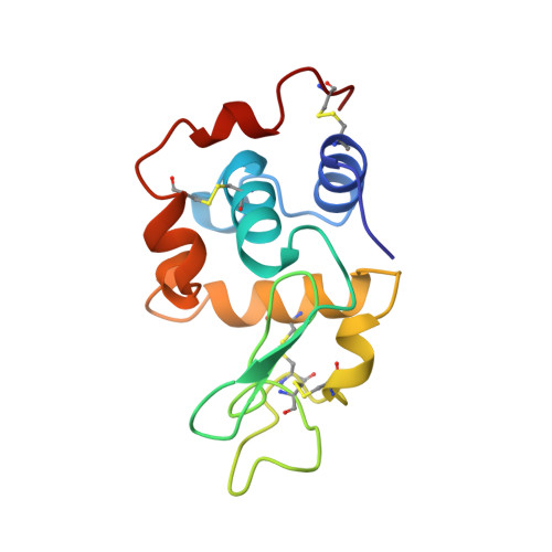 | |
UniProt | |||||
Entity Groups | |||||
| Sequence Clusters | 30% Identity50% Identity70% Identity90% Identity95% Identity100% Identity | ||||
| UniProt Group | P00698 | ||||
Sequence AnnotationsExpand | |||||
Reference Sequence | |||||
| Length ( Å ) | Angle ( ˚ ) |
|---|---|
| a = 79.1 | α = 90 |
| b = 79.1 | β = 90 |
| c = 37.9 | γ = 90 |
| Software Name | Purpose |
|---|---|
| CNS | refinement |


