2C9J | pdb_00002c9j

Structure of the fluorescent protein cmFP512 at 1.35A from Cerianthus membranaceus


Experimental Data Snapshot

  • Method: X-RAY DIFFRACTION
  • Resolution: 1.35 Å
  • R-Value Free: 
    0.255 (Depositor), 0.250 (DCC) 
  • R-Value Work: 
    0.228 (Depositor), 0.224 (DCC) 
  • R-Value Observed: 
    0.229 (Depositor) 

wwPDB Validation 3D Report Full Report

Validation slider image for 2C9J

This is version 2.1 of the entry. See complete history

Literature

Exploring Chromophore-Protein Interactions in Fluorescent Protein Cmfp512 from Cerianthus Membranaceus: X-Ray Structure Analysis and Optical Spectroscopy.

Nienhaus, K.Renzi, F.Vallone, B.Wiedenmann, J.Nienhaus, G.U.

(2006) Biochemistry 45: 12492

  • DOI: https://doi.org/10.1021/bi060885c
  • Primary Citation Related Structures: 
    2C9J

  • PubMed Abstract: 

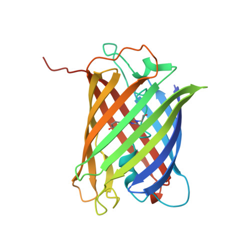
    Autofluorescent proteins of the GFP family all share the same three-dimensional beta-can fold; yet they exhibit widely different optical properties, arising either from chemical modification of the chromophore itself or from specific interactions of the chromophore with the surrounding protein moiety. Here we present a structural and spectroscopic characterization of the green fluorescent protein cmFP512 from Cerianthus membranaceus, a nonbioluminescent, azooxanthellate cnidarian, which has only approximately 22% sequence identity with Aequorea victoria GFP. The X-ray structure, obtained by molecular replacement at a resolution of 1. 35 A, shows the chromophore, formed from the tripeptide Gln-Tyr-Gly, in a hydrogen-bonded cage in the center of an 11-stranded beta-barrel, tightly restrained by adjacent residues and structural water molecules. It exists in a neutral (A) and an anionic (B) species, with absorption/emission maxima at 392/460 (pH 5) and 503/512 nm (pH 7). Their fractional populations and peak positions depend sensitively on pH, reflecting protonation of groups adjacent to the chromophore. The pH dependence of the spectra is explained by a protonation mechanism involving a hydrogen-bonded cluster of charged/polar groups. Cryospectroscopy at 12 K was also performed to analyze the vibronic coupling of the electronic transitions.


  • Organizational Affiliation
    • Department of Biophysics, University of Ulm, 89069 Ulm, Germany.

Macromolecule Content 

  • Total Structure Weight: 202.68 kDa 
  • Atom Count: 15,518 
  • Modeled Residue Count: 1,740 
  • Deposited Residue Count: 1,784 
  • Unique protein chains: 1

Macromolecules

Find similar proteins by:|  3D Structure
Entity ID: 1
MoleculeChains  Sequence LengthOrganismDetailsImage
GREEN FLUORESCENT PROTEIN FP512
A, B, C, D, E
A, B, C, D, E, F, G, H
223Cerianthus membranaceusMutation(s): 1 
UniProt
Find proteins for Q5ZQQ5 (Cerianthus membranaceus)
Explore Q5ZQQ5 
Go to UniProtKB:  Q5ZQQ5
Entity Groups
Sequence Clusters30% Identity50% Identity70% Identity90% Identity95% Identity100% Identity
UniProt GroupQ5ZQQ5
Sequence Annotations
Expand
Reference Sequence

Small Molecules

Modified Residues  1 Unique
IDChains TypeFormula2D DiagramParent
CRQ
Query on CRQ
A, B, C, D, E
A, B, C, D, E, F, G, H
L-PEPTIDE LINKINGC16 H16 N4 O5GLN, TYR, GLY

Experimental Data & Validation

Experimental Data

  • Method: X-RAY DIFFRACTION
  • Resolution: 1.35 Å
  • R-Value Free:  0.255 (Depositor), 0.250 (DCC) 
  • R-Value Work:  0.228 (Depositor), 0.224 (DCC) 
  • R-Value Observed: 0.229 (Depositor) 
Space Group: P 1
Unit Cell:
Length ( Å )Angle ( ˚ )
a = 54.001α = 83.8
b = 60.108β = 89.98
c = 125.4γ = 73.85
Software Package:
Software NamePurpose
REFMACrefinement
DENZOdata reduction
SCALEPACKdata scaling
MOLREPphasing

Structure Validation

View Full Validation Report


Entry History 

Deposition Data

Revision History  (Full details and data files)

  • Version 1.0: 2006-10-30
    Type: Initial release
  • Version 1.1: 2011-05-08
    Changes: Version format compliance
  • Version 1.2: 2011-07-13
    Changes: Version format compliance
  • Version 1.3: 2019-10-23
    Changes: Data collection, Database references, Derived calculations, Other
  • Version 2.0: 2023-11-15
    Changes: Advisory, Atomic model, Data collection, Database references, Derived calculations
  • Version 2.1: 2024-11-13
    Changes: Structure summary