2C9I | pdb_00002c9i

Structure of the fluorescent protein asFP499 from Anemonia sulcata


Experimental Data Snapshot

  • Method: X-RAY DIFFRACTION
  • Resolution: 1.82 Å
  • R-Value Free: 
    0.292 (Depositor), 0.290 (DCC) 
  • R-Value Work: 
    0.245 (Depositor), 0.244 (DCC) 
  • R-Value Observed: 
    0.248 (Depositor) 

wwPDB Validation 3D Report Full Report

Validation slider image for 2C9I

This is version 2.1 of the entry. See complete history

Literature

Chromophore-Protein Interactions in the Anthozoan Green Fluorescent Protein Asfp499

Nienhaus, K.Renzi, F.Vallone, B.Wiedenmann, J.Nienhaus, G.U.

(2006) Biophys J 91: 4210

  • DOI: https://doi.org/10.1529/biophysj.106.087411
  • Primary Citation Related Structures: 
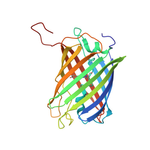
    2C9I

  • PubMed Abstract: 

    Despite their similar fold topologies, anthozoan fluorescent proteins (FPs) can exhibit widely different optical properties, arising either from chemical modification of the chromophore itself or from specific interactions of the chromophore with the surrounding protein moiety. Here we present a structural and spectroscopic investigation of the green FP asFP499 from the sea anemone Anemonia sulcata var. rufescens to explore the effects of the protein environment on the chromophore. The optical absorption and fluorescence spectra reveal two discrete species populated in significant proportions over a wide pH range. Moreover, multiple protonation reactions are evident from the observed pH-dependent spectral changes. The x-ray structure of asFP499, determined by molecular replacement at a resolution of 1.85 A, shows the typical beta-barrel fold of the green FP from Aequorea victoria (avGFP). In its center, the chromophore, formed from the tripeptide Gln(63)-Tyr(64)-Gly(65), is tightly held by multiple hydrogen bonds in a polar cage that is structurally quite dissimilar to that of avGFP. The x-ray structure provides interesting clues as to how the spectroscopic properties are fine tuned by the chromophore environment.


  • Organizational Affiliation
    • Department of Biophysics, University of Ulm, 89069 Ulm, Germany. uli@uiuc.edu

Macromolecule Content 

  • Total Structure Weight: 203.03 kDa 
  • Atom Count: 15,798 
  • Modeled Residue Count: 1,805 
  • Deposited Residue Count: 1,808 
  • Unique protein chains: 1

Macromolecules

Find similar proteins by:|  3D Structure
Entity ID: 1
MoleculeChains  Sequence LengthOrganismDetailsImage
GREEN FLUORESCENT PROTEIN ASFP499
A, B, C, D, E
A, B, C, D, E, F, G, H
226Anemonia sulcataMutation(s): 1 
UniProt
Find proteins for Q9GPI6 (Anemonia sulcata)
Explore Q9GPI6 
Go to UniProtKB:  Q9GPI6
Entity Groups
Sequence Clusters30% Identity50% Identity70% Identity90% Identity95% Identity100% Identity
UniProt GroupQ9GPI6
Sequence Annotations
Expand
Reference Sequence

Small Molecules

Modified Residues  1 Unique
IDChains TypeFormula2D DiagramParent
CRQ
Query on CRQ
A, B, C, D, E
A, B, C, D, E, F, G, H
L-PEPTIDE LINKINGC16 H16 N4 O5GLN, TYR, GLY

Experimental Data & Validation

Experimental Data

  • Method: X-RAY DIFFRACTION
  • Resolution: 1.82 Å
  • R-Value Free:  0.292 (Depositor), 0.290 (DCC) 
  • R-Value Work:  0.245 (Depositor), 0.244 (DCC) 
  • R-Value Observed: 0.248 (Depositor) 
Space Group: P 1 21 1
Unit Cell:
Length ( Å )Angle ( ˚ )
a = 71.88α = 90
b = 135.126β = 106.93
c = 95.071γ = 90
Software Package:
Software NamePurpose
REFMACrefinement
DENZOdata reduction
SCALEPACKdata scaling
AMoREphasing

Structure Validation

View Full Validation Report


Entry History 

Deposition Data

Revision History  (Full details and data files)

  • Version 1.0: 2007-01-16
    Type: Initial release
  • Version 1.1: 2011-05-08
    Changes: Version format compliance
  • Version 1.2: 2011-07-13
    Changes: Version format compliance
  • Version 1.3: 2019-10-23
    Changes: Data collection, Database references, Derived calculations, Other
  • Version 2.0: 2023-11-15
    Changes: Advisory, Atomic model, Data collection, Database references, Derived calculations
  • Version 2.1: 2024-10-16
    Changes: Structure summary