2C91

mouse succinic semialdehyde reductase, AKR7A5


Experimental Data Snapshot

  • Method: X-RAY DIFFRACTION
  • Resolution: 2.30 Å
  • R-Value Free: 0.208 
  • R-Value Work: 0.160 
  • R-Value Observed: 0.162 

wwPDB Validation   3D Report Full Report


Ligand Structure Quality Assessment 


This is version 1.3 of the entry. See complete history


Literature

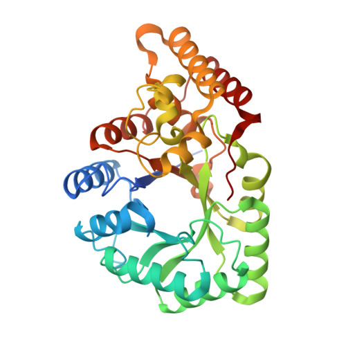
Crystal Structure of Mouse Succinic Semialdehyde Reductase Akr7A5: Structural Basis for Substrate Specificity.

Zhu, X.Lapthorn, A.J.Ellis, E.M.

(2006) Biochemistry 45: 1562

  • DOI: https://doi.org/10.1021/bi051610k
  • Primary Citation of Related Structures:  
    2C91

  • PubMed Abstract: 

    The aldo-keto reductases make up a superfamily of enzymes which can reduce a variety of aldehydes and ketones to their corresponding alcohols. Within each family are distinct preferences for certain substrates, presumably reflecting their role within the cell. The original member of the AKR7A subfamily was purified from liver as an aflatoxin dialdehyde reductase AKR7A1. However, recent additions to the family have revealed that even closely related enzymes have clear substrate preferences with AKR7A2, AKR7A4, and AKR7A5 showing much higher affinities for succinic semialdehyde (SSA) than does AKR7A1. To investigate the structural basis of this specificity, the crystal structure of mouse AKR7A5 has been determined to better than 2.5 A resolution. The structure is of the ternary complex of the enzyme with NADP+ and tartrate as an inhibitor. This structure has the same overall fold as the previously determined structure of AKR7A1; however, there are a number of differences in loops around the active site that contribute to observed differences in the substrate specificity between the AKR7A enzymes. Several differences are the result of bulky hydrophobic residues found in AKR7A5, namely, Met44, Trp77, and Trp224, which significantly restrict the size and modify the architecture of the substrate-binding pocket, producing a tighter or less flexible binding site for SSA than in AKR7A1. Site-directed mutagenesis was used to introduce Met44, Trp77, and Trp224 individually into AKR7A1, to test if they improved the affinity of the enzyme for SSA. Each mutation showed improved affinity for SSA, with Trp77Met having the largest effect. This confirms the role of these amino acids as substrate determinants for SSA.


  • Organizational Affiliation

    Department of Chemistry, University of Glasgow, Glasgow G12 8QQ, UK.


Macromolecules
Find similar proteins by:  (by identity cutoff)  |  3D Structure
Entity ID: 1
MoleculeChains Sequence LengthOrganismDetailsImage
AFLATOXIN B1 ALDEHYDE REDUCTASE MEMBER 2
A, B, C, D, E
A, B, C, D, E, F, G, H, I, J
338Mus musculusMutation(s): 0 
EC: 1
UniProt & NIH Common Fund Data Resources
Find proteins for Q8CG76 (Mus musculus)
Explore Q8CG76 
Go to UniProtKB:  Q8CG76
IMPC:  MGI:107796
Entity Groups  
Sequence Clusters30% Identity50% Identity70% Identity90% Identity95% Identity100% Identity
UniProt GroupQ8CG76
Sequence Annotations
Expand
  • Reference Sequence
Small Molecules
Ligands 5 Unique
IDChains Name / Formula / InChI Key2D Diagram3D Interactions
NAP
Query on NAP

Download Ideal Coordinates CCD File 
BA [auth D]
DB [auth J]
HA [auth E]
K [auth A]
MA [auth F]
BA [auth D],
DB [auth J],
HA [auth E],
K [auth A],
MA [auth F],
P [auth B],
PA [auth G],
UA [auth H],
V [auth C],
YA [auth I]
NADP NICOTINAMIDE-ADENINE-DINUCLEOTIDE PHOSPHATE
C21 H28 N7 O17 P3
XJLXINKUBYWONI-NNYOXOHSSA-N
MES
Query on MES

Download Ideal Coordinates CCD File 
AA [auth C],
CB [auth I],
GA [auth D],
JB [auth J],
U [auth B]
2-(N-MORPHOLINO)-ETHANESULFONIC ACID
C6 H13 N O4 S
SXGZJKUKBWWHRA-UHFFFAOYSA-N
TLA
Query on TLA

Download Ideal Coordinates CCD File 
CA [auth D]
EB [auth J]
IA [auth E]
L [auth A]
NA [auth F]
CA [auth D],
EB [auth J],
IA [auth E],
L [auth A],
NA [auth F],
Q [auth B],
QA [auth G],
VA [auth H],
W [auth C],
ZA [auth I]
L(+)-TARTARIC ACID
C4 H6 O6
FEWJPZIEWOKRBE-JCYAYHJZSA-N
PO4
Query on PO4

Download Ideal Coordinates CCD File 
IB [auth J],
O [auth A]
PHOSPHATE ION
O4 P
NBIIXXVUZAFLBC-UHFFFAOYSA-K
GOL
Query on GOL

Download Ideal Coordinates CCD File 
AB [auth I]
BB [auth I]
DA [auth D]
EA [auth D]
FA [auth D]
AB [auth I],
BB [auth I],
DA [auth D],
EA [auth D],
FA [auth D],
FB [auth J],
GB [auth J],
HB [auth J],
JA [auth E],
KA [auth E],
LA [auth E],
M [auth A],
N [auth A],
OA [auth F],
R [auth B],
RA [auth G],
S [auth B],
SA [auth G],
T [auth B],
TA [auth G],
WA [auth H],
X [auth C],
XA [auth H],
Y [auth C],
Z [auth C]
GLYCEROL
C3 H8 O3
PEDCQBHIVMGVHV-UHFFFAOYSA-N
Experimental Data & Validation

Experimental Data

  • Method: X-RAY DIFFRACTION
  • Resolution: 2.30 Å
  • R-Value Free: 0.208 
  • R-Value Work: 0.160 
  • R-Value Observed: 0.162 
  • Space Group: P 1
Unit Cell:
Length ( Å )Angle ( ˚ )
a = 98.532α = 90.02
b = 159.238β = 119.4
c = 96.698γ = 78.5
Software Package:
Software NamePurpose
REFMACrefinement
DENZOdata reduction
SCALEPACKdata scaling
AMoREphasing

Structure Validation

View Full Validation Report


Ligand Structure Quality Assessment 


Entry History 

Deposition Data

Revision History  (Full details and data files)

  • Version 1.0: 2006-02-15
    Type: Initial release
  • Version 1.1: 2011-07-13
    Changes: Advisory, Version format compliance
  • Version 1.2: 2019-05-08
    Changes: Data collection, Experimental preparation, Other
  • Version 1.3: 2023-12-13
    Changes: Data collection, Database references, Derived calculations, Other, Refinement description