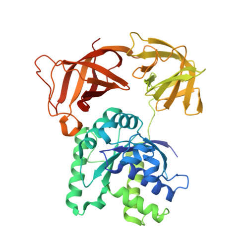
2C78

EF-Tu complexed with a GTP analog and the antibiotic pulvomycin


Experimental Data Snapshot

  • Method: X-RAY DIFFRACTION
  • Resolution: 1.40 Å
  • R-Value Free: 0.216 
  • R-Value Work: 0.197 
  • R-Value Observed: 0.197 

Starting Model: experimental
View more details

wwPDB Validation   3D Report Full Report


Ligand Structure Quality Assessment 


This is version 1.5 of the entry. See complete history


Literature

Structural basis of the action of pulvomycin and GE2270 A on elongation factor Tu.

Parmeggiani, A.Krab, I.M.Okamura, S.Nielsen, R.C.Nyborg, J.Nissen, P.

(2006) Biochemistry 45: 6846-6857

  • DOI: https://doi.org/10.1021/bi0525122
  • Primary Citation of Related Structures:  
    2C77, 2C78

  • PubMed Abstract: 

    Pulvomycin inhibits protein synthesis by preventing the formation of the ternary complex between elongation factor Tu (EF-Tu) x GTP and aa-tRNA. In this work, the crystal structure of Thermus thermophilus EF-Tu x pulvomycin in complex with the GTP analogue guanylyl imino diphosphate (GDPNP) at 1.4 A resolution reveals an antibiotic binding site extending from the domain 1-3 interface to domain 2, overlapping the domain 1-2-3 junction. Pulvomycin binding interferes with the binding of the 3'-aminoacyl group, the acceptor stem, and 5' end of tRNA. Only part of pulvomycin overlaps the binding site of GE2270 A, a domain 2-bound antibiotic of a structure unrelated to pulvomycin, which also hinders aa-tRNA binding. The structure of the T. thermophilus EF-Tu x GDPNP x GE2270 A complex at 1.6 A resolution shows that GE2270 A interferes with the binding of the 3'-aminoacyl group and part of the acceptor stem of aa-tRNA but not with the 5' end. Both compounds, pulvomycin more markedly, hinder the correct positioning of domain 1 over domains 2 and 3 that characterizes the active form of EF-Tu, while they affect the domain 1 switch regions that control the EF-Tu x GDP/GTP transitions in different ways. This work reveals how two antibiotics with different structures and binding modes can employ a similar mechanism of action.


  • Organizational Affiliation

    Department of Molecular Biology, University of Aarhus, Gustav Wieds Vej 10 C, DK-8000 Aarhus C, Denmark. andrea@bioxray.dk


Macromolecules
Find similar proteins by:  (by identity cutoff)  |  3D Structure
Entity ID: 1
MoleculeChains Sequence LengthOrganismDetailsImage
ELONGATION FACTOR TU-A405Thermus thermophilus HB8Mutation(s): 0 
EC: 3.1.5.1
UniProt
Find proteins for Q5SHN6 (Thermus thermophilus (strain ATCC 27634 / DSM 579 / HB8))
Explore Q5SHN6 
Go to UniProtKB:  Q5SHN6
Entity Groups  
Sequence Clusters30% Identity50% Identity70% Identity90% Identity95% Identity100% Identity
UniProt GroupQ5SHN6
Sequence Annotations
Expand
  • Reference Sequence
Small Molecules
Ligands 3 Unique
IDChains Name / Formula / InChI Key2D Diagram3D Interactions
PUL
Query on PUL

Download Ideal Coordinates CCD File 
D [auth A](1S,2S,3E,5E,7E,10S,11S,12S)-12-[(2R,4E,6E,8Z,10R,12E,14E,16Z,18S,19Z)-10,18-DIHYDROXY-12,16,19-TRIMETHYL-11,22-DIOXOOX ACYCLODOCOSA-4,6,8,12,14,16,19-HEPTAEN-2-YL]-2,11-DIHYDROXY-1,10-DIMETHYL-9-OXOTRIDECA-3,5,7-TRIEN-1-YL 6-DEOXY-2,4-DI-O-METHYL-BETA-L-GALACTOPYRANOSIDE
C47 H66 O13
FXSFWUNCIOIMAC-VXILMCRLSA-N
GNP
Query on GNP

Download Ideal Coordinates CCD File 
B [auth A]PHOSPHOAMINOPHOSPHONIC ACID-GUANYLATE ESTER
C10 H17 N6 O13 P3
UQABYHGXWYXDTK-UUOKFMHZSA-N
MG
Query on MG

Download Ideal Coordinates CCD File 
C [auth A]MAGNESIUM ION
Mg
JLVVSXFLKOJNIY-UHFFFAOYSA-N
Experimental Data & Validation

Experimental Data

  • Method: X-RAY DIFFRACTION
  • Resolution: 1.40 Å
  • R-Value Free: 0.216 
  • R-Value Work: 0.197 
  • R-Value Observed: 0.197 
  • Space Group: P 65
Unit Cell:
Length ( Å )Angle ( ˚ )
a = 86.686α = 90
b = 86.686β = 90
c = 105.514γ = 120
Software Package:
Software NamePurpose
CNSrefinement
DENZOdata reduction
SCALEPACKdata scaling
AMoREphasing

Structure Validation

View Full Validation Report


Ligand Structure Quality Assessment 


Entry History 

Deposition Data

Revision History  (Full details and data files)

  • Version 1.0: 2006-03-16
    Type: Initial release
  • Version 1.1: 2011-05-08
    Changes: Version format compliance
  • Version 1.2: 2011-07-13
    Changes: Version format compliance
  • Version 1.3: 2018-03-28
    Changes: Database references, Source and taxonomy
  • Version 1.4: 2019-07-24
    Changes: Data collection
  • Version 1.5: 2023-12-13
    Changes: Data collection, Database references, Derived calculations, Other, Refinement description, Structure summary