2C1D | pdb_00002c1d

Crystal structure of SoxXA from P. pantotrophus


Experimental Data Snapshot

  • Method: X-RAY DIFFRACTION
  • Resolution: 1.92 Å
  • R-Value Free: 
    0.211 (Depositor) 
  • R-Value Work: 
    0.159 (Depositor), 0.174 (DCC) 
  • R-Value Observed: 
    0.161 (Depositor) 

Starting Model: experimental
View more details

wwPDB Validation 3D Report Full Report

Validation slider image for 2C1D

Ligand Structure Quality Assessment 


This is version 1.4 of the entry. See complete history

Literature

Structure of the Cytochrome Complex Soxxa of Paracoccus Pantotrophus, a Heme Enzyme Initiating Chemotrophic Sulfur Oxidation.

Dambe, T.Quentmeier, A.Rother, D.Friedrich, C.Scheidig, A.J.

(2005) J Struct Biol 152: 229

  • DOI: https://doi.org/10.1016/j.jsb.2005.09.002
  • Primary Citation Related Structures: 
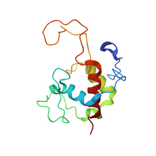
    2C1D

  • PubMed Abstract: 

    The sulfur-oxidizing enzyme system (Sox) of the chemotroph Paracoccus pantotrophus is composed of several proteins, which together oxidize hydrogen sulfide, sulfur, thiosulfate or sulfite and transfers the gained electrons to the respiratory chain. The hetero-dimeric cytochrome c complex SoxXA functions as heme enzyme and links covalently the sulfur substrate to the thiol of the cysteine-138 residue of the SoxY protein of the SoxYZ complex. Here, we report the crystal structure of the c-type cytochrome complex SoxXA. The structure could be solved by molecular replacement and refined to a resolution of 1.9A identifying the axial heme-iron coordination involving an unusual Cys-251 thiolate of heme2. Distance measurements between the three heme groups provide deeper insight into the electron transport inside SoxXA and merge in a better understanding of the initial step of the aerobic sulfur oxidation process in chemotrophic bacteria.


  • Organizational Affiliation
    • Max-Planck-Institut für Molekulare Physiologie, Abteilung Physikalische Biochemie, Otto-Hahn-Strasse 11, D-44225 Dortmund, Germany.

Macromolecule Content 

  • Total Structure Weight: 182.66 kDa 
  • Atom Count: 14,878 
  • Modeled Residue Count: 1,604 
  • Deposited Residue Count: 1,604 
  • Unique protein chains: 2

Macromolecules

Find similar proteins by:|  3D Structure
Entity ID: 1
MoleculeChains  Sequence LengthOrganismDetailsImage
SOXA
A, C, E, G
264Paracoccus denitrificansMutation(s): 0 
EC: 2.8.5.2
UniProt
Find proteins for O33434 (Paracoccus pantotrophus)
Explore O33434 
Go to UniProtKB:  O33434
Entity Groups
Sequence Clusters30% Identity50% Identity70% Identity90% Identity95% Identity100% Identity
UniProt GroupO33434
Sequence Annotations
Expand
Reference Sequence
Find similar proteins by:|  3D Structure
Entity ID: 2
MoleculeChains  Sequence LengthOrganismDetailsImage
SOXX
B, D, F, H
137Paracoccus pantotrophusMutation(s): 0 
UniProt
Find proteins for Q9LCV0 (Paracoccus denitrificans)
Explore Q9LCV0 
Go to UniProtKB:  Q9LCV0
Entity Groups
Sequence Clusters30% Identity50% Identity70% Identity90% Identity95% Identity100% Identity
UniProt GroupQ9LCV0
Sequence Annotations
Expand
Reference Sequence

Small Molecules

Ligands 2 Unique
IDChains Name / Formula / InChI Key2D Diagram3D Interactions
HEC

Query on HEC


Download:Ideal Coordinates CCD File
AA [auth G]
BA [auth G]
FA [auth H]
I [auth A]
J [auth A]
AA [auth G],
BA [auth G],
FA [auth H],
I [auth A],
J [auth A],
O [auth B],
P [auth C],
Q [auth C],
U [auth D],
V [auth E],
W [auth E],
Z [auth F]
HEME C
C34 H34 Fe N4 O4
HXQIYSLZKNYNMH-LJNAALQVSA-N
ZN

Query on ZN


Download:Ideal Coordinates CCD File
CA [auth G]
DA [auth G]
EA [auth G]
K [auth A]
L [auth A]
CA [auth G],
DA [auth G],
EA [auth G],
K [auth A],
L [auth A],
M [auth A],
N [auth A],
R [auth C],
S [auth C],
T [auth C],
X [auth E],
Y [auth E]
ZINC ION
Zn
PTFCDOFLOPIGGS-UHFFFAOYSA-N
Modified Residues  1 Unique
IDChains TypeFormula2D DiagramParent
CSS
Query on CSS
A, C, E, G
L-PEPTIDE LINKINGC3 H7 N O2 S2CYS

Experimental Data & Validation

Experimental Data

  • Method: X-RAY DIFFRACTION
  • Resolution: 1.92 Å
  • R-Value Free:  0.211 (Depositor) 
  • R-Value Work:  0.159 (Depositor), 0.174 (DCC) 
  • R-Value Observed: 0.161 (Depositor) 
Space Group: P 1 21 1
Unit Cell:
Length ( Å )Angle ( ˚ )
a = 42.86α = 90
b = 180.01β = 92.83
c = 117.93γ = 90
Software Package:
Software NamePurpose
REFMACrefinement
XDSdata reduction
XSCALEdata scaling
MOLREPphasing

Structure Validation

View Full Validation Report


Ligand Structure Quality Assessment 


Entry History 

Deposition Data

Revision History  (Full details and data files)

  • Version 1.0: 2005-10-06
    Type: Initial release
  • Version 1.1: 2011-05-08
    Changes: Version format compliance
  • Version 1.2: 2011-07-13
    Changes: Version format compliance
  • Version 1.3: 2023-12-13
    Changes: Data collection, Database references, Derived calculations, Other, Refinement description
  • Version 1.4: 2024-10-16
    Changes: Structure summary