2BT4 | pdb_00002bt4

Type II Dehydroquinase inhibitor complex


Experimental Data Snapshot

  • Method: X-RAY DIFFRACTION
  • Resolution: 1.70 Å
  • R-Value Free: 
    0.248 (Depositor) 
  • R-Value Work: 
    0.197 (Depositor) 
  • R-Value Observed: 
    0.199 (Depositor) 

Starting Model: experimental
View more details

wwPDB Validation 3D Report Full Report

Validation slider image for 2BT4

This is version 1.3 of the entry. See complete history

Literature

Rational Design of New Bifunctional Inhibitors of Type II Dehydroquinase.

Toscano, M.D.Stewart, K.A.Coggins, J.R.Lapthorn, A.J.Abell, C.

(2005) Org Biomol Chem 3: 3102

  • DOI: https://doi.org/10.1039/b507156a
  • Primary Citation Related Structures: 
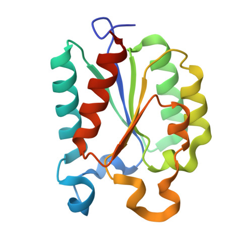
    2BT4

  • PubMed Abstract: 

    Selective inhibitors of type II dehydroquinase were rationally designed to explore a second binding-pocket in the active-site. The molecular modelling, synthesis, inhibition studies and crystal structure determination are described.


  • Organizational Affiliation
    • University Chemical Laboratory, Lensfield Road, Cambridge CB2 1EW, UK.

Macromolecule Content 

  • Total Structure Weight: 206.11 kDa 
  • Atom Count: 15,641 
  • Modeled Residue Count: 1,788 
  • Deposited Residue Count: 1,884 
  • Unique protein chains: 1

Macromolecules

Find similar proteins by:|  3D Structure
Entity ID: 1
MoleculeChains  Sequence LengthOrganismDetailsImage
3-DEHYDROQUINATE DEHYDRATASE
A, B, C, D, E
A, B, C, D, E, F, G, H, I, J, K, L
157Streptomyces coelicolorMutation(s): 0 
EC: 4.2.1.10
UniProt
Find proteins for P15474 (Streptomyces coelicolor (strain ATCC BAA-471 / A3(2) / M145))
Explore P15474 
Go to UniProtKB:  P15474
Entity Groups
Sequence Clusters30% Identity50% Identity70% Identity90% Identity95% Identity100% Identity
UniProt GroupP15474
Sequence Annotations
Expand
Reference Sequence

Small Molecules

Ligands 4 Unique
IDChains Name / Formula / InChI Key2D Diagram3D Interactions
CA2

Query on CA2


Download:Ideal Coordinates CCD File
AA [auth F]
DA [auth G]
FA [auth H]
IA [auth I]
LA [auth J]
AA [auth F],
DA [auth G],
FA [auth H],
IA [auth I],
LA [auth J],
M [auth A],
NA [auth K],
PA [auth L],
Q [auth B],
S [auth C],
U [auth D],
X [auth E]
(1S,3R,4R,5S)-1,3,4-TRIHYDROXY-5-(3-PHENOXYPROPYL)CYCLOHEXANECARBOXYLIC ACID
C16 H22 O6
SCUFESRLGCQXRX-DCDXPUDHSA-N
TRS

Query on TRS


Download:Ideal Coordinates CCD File
BA [auth F],
GA [auth H],
O [auth A],
RA [auth L]
2-AMINO-2-HYDROXYMETHYL-PROPANE-1,3-DIOL
C4 H12 N O3
LENZDBCJOHFCAS-UHFFFAOYSA-O
PO4

Query on PO4


Download:Ideal Coordinates CCD File
JA [auth I],
N [auth A],
QA [auth L],
V [auth D]
PHOSPHATE ION
O4 P
NBIIXXVUZAFLBC-UHFFFAOYSA-K
GOL

Query on GOL


Download:Ideal Coordinates CCD File
CA [auth F]
EA [auth G]
HA [auth H]
KA [auth I]
MA [auth J]
CA [auth F],
EA [auth G],
HA [auth H],
KA [auth I],
MA [auth J],
OA [auth K],
P [auth A],
R [auth B],
T [auth C],
W [auth D],
Y [auth E],
Z [auth E]
GLYCEROL
C3 H8 O3
PEDCQBHIVMGVHV-UHFFFAOYSA-N

Experimental Data & Validation

Experimental Data

  • Method: X-RAY DIFFRACTION
  • Resolution: 1.70 Å
  • R-Value Free:  0.248 (Depositor) 
  • R-Value Work:  0.197 (Depositor) 
  • R-Value Observed: 0.199 (Depositor) 
Space Group: P 1
Unit Cell:
Length ( Å )Angle ( ˚ )
a = 196.616α = 65.91
b = 196.487β = 65.91
c = 240.626γ = 90.01
Software Package:
Software NamePurpose
REFMACrefinement
DENZOdata reduction
SCALEPACKdata scaling
AMoREphasing

Structure Validation

View Full Validation Report


Entry History 

Deposition Data

Revision History  (Full details and data files)

  • Version 1.0: 2006-02-13
    Type: Initial release
  • Version 1.1: 2011-06-09
    Changes: Version format compliance
  • Version 1.2: 2011-07-13
    Changes: Version format compliance
  • Version 1.3: 2023-12-13
    Changes: Data collection, Database references, Derived calculations, Refinement description