2BOB

Potassium channel KcsA-Fab complex in thallium with tetrabutylammonium (TBA)


Experimental Data Snapshot

  • Method: X-RAY DIFFRACTION
  • Resolution: 2.76 Å
  • R-Value Free: 0.251 
  • R-Value Work: 0.220 
  • R-Value Observed: 0.222 

wwPDB Validation   3D Report Full Report


Ligand Structure Quality Assessment 


This is version 1.3 of the entry. See complete history


Literature

Structural Basis of Tea Blockade in a Model Potassium Channel

Lenaeus, M.J.Vamvouka, M.Focia, P.J.Gross, A.

(2005) Nat Struct Mol Biol 12: 454

  • DOI: https://doi.org/10.1038/nsmb929
  • Primary Citation of Related Structures:  
    2BOB, 2BOC

  • PubMed Abstract: 
  • Potassium channels catalyze the selective transfer of potassium across the cell membrane and are essential for setting the resting potential in cells, controlling heart rate and modulating the firing pattern in neurons. Tetraethylammonium (TEA) blocks ion conduction through potassium channels in a voltage-dependent manner from both sides of the membrane ...

    Potassium channels catalyze the selective transfer of potassium across the cell membrane and are essential for setting the resting potential in cells, controlling heart rate and modulating the firing pattern in neurons. Tetraethylammonium (TEA) blocks ion conduction through potassium channels in a voltage-dependent manner from both sides of the membrane. Here we show the structural basis of TEA blockade by cocrystallizing the prokaryotic potassium channel KcsA with two selective TEA analogs. TEA binding at both sites alters ion occupancy in the selectivity filter; these findings underlie the mutual destabilization and voltage-dependence of TEA blockade. We propose that TEA blocks potassium channels by acting as a potassium analog at the dehydration transition step during permeation.


    Organizational Affiliation

    Department of Molecular Pharmacology and Biological Chemistry, Northwestern University Medical School, 303 East Chicago Avenue, Chicago, Illinois 60611, USA.


Macromolecules
Find similar proteins by:  (by identity cutoff)  |  3D Structure
Entity ID: 1
MoleculeChainsSequence LengthOrganismDetailsImage
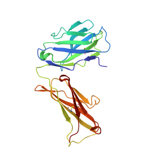
ANTIBODY FAB FRAGMENT HEAVY CHAINA219Mus musculusMutation(s): 0 
Entity Groups  
Sequence Clusters30% Identity50% Identity70% Identity90% Identity95% Identity100% Identity
Protein Feature View
Expand
  • Reference Sequence
Find similar proteins by:  (by identity cutoff)  |  3D Structure
Entity ID: 2
MoleculeChainsSequence LengthOrganismDetailsImage
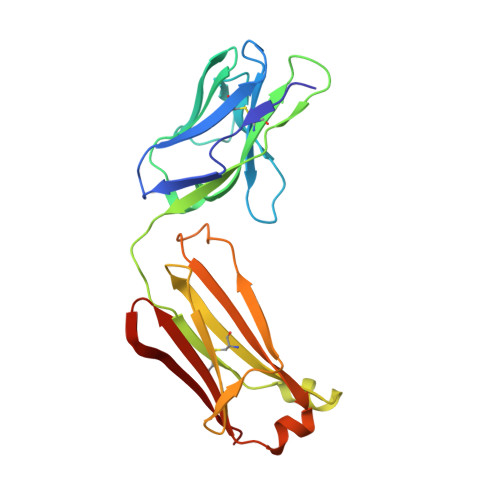
ANTIBODY FAB FRAGMENT LIGHT CHAINB212Mus musculusMutation(s): 0 
Entity Groups  
Sequence Clusters30% Identity50% Identity70% Identity90% Identity95% Identity100% Identity
Protein Feature View
Expand
  • Reference Sequence
Find similar proteins by:  (by identity cutoff)  |  3D Structure
Entity ID: 3
MoleculeChainsSequence LengthOrganismDetailsImage
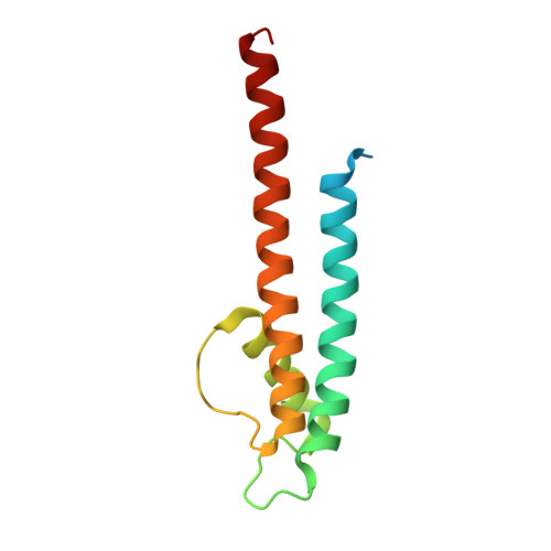
POTASSIUM CHANNEL KCSAC124Streptomyces lividansMutation(s): 1 
Gene Names: kcsAskc1
Membrane Entity: Yes 
UniProt
Find proteins for P0A334 (Streptomyces lividans)
Explore P0A334 
Go to UniProtKB:  P0A334
Entity Groups  
Sequence Clusters30% Identity50% Identity70% Identity90% Identity95% Identity100% Identity
UniProt GroupP0A334
Protein Feature View
Expand
  • Reference Sequence
Small Molecules
Ligands 3 Unique
IDChainsName / Formula / InChI Key2D Diagram3D Interactions
TBA
Query on TBA

Download Ideal Coordinates CCD File 
F [auth C]TETRABUTYLAMMONIUM ION
C16 H36 N
DZLFLBLQUQXARW-UHFFFAOYSA-N
 Ligand Interaction
TL
Query on TL

Download Ideal Coordinates CCD File 
D [auth C]THALLIUM (I) ION
Tl
ZLUSCZLCHQSJRU-UHFFFAOYSA-N
 Ligand Interaction
CO
Query on CO

Download Ideal Coordinates CCD File 
E [auth C]COBALT (II) ION
Co
XLJKHNWPARRRJB-UHFFFAOYSA-N
 Ligand Interaction
Experimental Data & Validation

Experimental Data

  • Method: X-RAY DIFFRACTION
  • Resolution: 2.76 Å
  • R-Value Free: 0.251 
  • R-Value Work: 0.220 
  • R-Value Observed: 0.222 
  • Space Group: I 4
Unit Cell:
Length ( Å )Angle ( ˚ )
a = 155.267α = 90
b = 155.267β = 90
c = 75.555γ = 90
Software Package:
Software NamePurpose
REFMACrefinement
XDSdata reduction
XDSdata scaling
CNSphasing

Structure Validation

View Full Validation Report


Ligand Structure Quality Assessment 


Entry History 

Deposition Data

Revision History  (Full details and data files)

  • Version 1.0: 2005-04-27
    Type: Initial release
  • Version 1.1: 2011-05-08
    Changes: Version format compliance
  • Version 1.2: 2011-07-13
    Changes: Version format compliance
  • Version 1.3: 2017-07-12
    Changes: Derived calculations