2BL0 | pdb_00002bl0

Physarum polycephalum myosin II regulatory domain


Experimental Data Snapshot

  • Method: X-RAY DIFFRACTION
  • Resolution: 1.75 Å
  • R-Value Free: 
    0.252 (Depositor), 0.261 (DCC) 
  • R-Value Work: 
    0.219 (Depositor), 0.223 (DCC) 
  • R-Value Observed: 
    0.221 (Depositor) 

wwPDB Validation 3D Report Full Report

Validation slider image for 2BL0

This is version 1.3 of the entry. See complete history

Literature

Structural Evidence for Non-Canonical Binding of Ca2+ to a Canonical EF-Hand of a Conventional Myosin.

Debreczeni, J.E.Farkas, L.Harmat, V.Hetenyi, C.Hajdu, I.Zavodszky, P.Kohama, K.Nyitray, L.

(2005) J Biological Chem 280: 41458

  • DOI: https://doi.org/10.1074/jbc.M506315200
  • Primary Citation Related Structures: 
    2BL0

  • PubMed Abstract: 

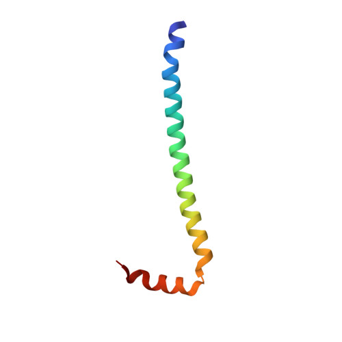
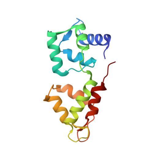
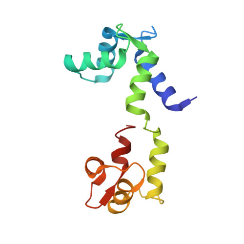
    We have previously identified a single inhibitory Ca2+-binding site in the first EF-hand of the essential light chain of Physarum conventional myosin (Farkas, L., Malnasi-Csizmadia, A., Nakamura, A., Kohama, K., and Nyitray, L. (2003) J. Biol. Chem. 278, 27399-27405). As a general rule, conformation of the EF-hand-containing domains in the calmodulin family is "closed" in the absence and "open" in the presence of bound cations; a notable exception is the unusual Ca2+-bound closed domain in the essential light chain of the Ca2+-activated scallop muscle myosin. Here we have reported the 1.8 A resolution structure of the regulatory domain (RD) of Physarum myosin II in which Ca2+ is bound to a canonical EF-hand that is also in a closed state. The 12th position of the EF-hand loop, which normally provides a bidentate ligand for Ca2+ in the open state, is too far in the structure to participate in coordination of the ion. The structure includes a second Ca2+ that only mediates crystal contacts. To reveal the mechanism behind the regulatory effect of Ca2+, we compared conformational flexibilities of the liganded and unliganded RD. Our working hypothesis, i.e. the modulatory effect of Ca2+ on conformational flexibility of RD, is in line with the observed suppression of hydrogen-deuterium exchange rate in the Ca2+-bound form, as well as with results of molecular dynamics calculations. Based on this evidence, we concluded that Ca2+-induced change in structural dynamics of RD is a major factor in Ca2+-mediated regulation of Physarum myosin II activity.


  • Organizational Affiliation
    • Department of Biochemistry, Eötvös Loránd University, Budapest H-1117, Hungary.

Macromolecule Content 

  • Total Structure Weight: 39.79 kDa 
  • Atom Count: 3,078 
  • Modeled Residue Count: 350 
  • Deposited Residue Count: 350 
  • Unique protein chains: 3

Macromolecules

Find similar proteins by:|  3D Structure
Entity ID: 1
MoleculeChains  Sequence LengthOrganismDetailsImage
MAJOR PLASMODIAL MYOSIN HEAVY CHAIN63Badhamia polycephalaMutation(s): 0 
UniProt
Find proteins for Q9BJD3 (Physarum polycephalum)
Explore Q9BJD3 
Go to UniProtKB:  Q9BJD3
Entity Groups
Sequence Clusters30% Identity50% Identity70% Identity90% Identity95% Identity100% Identity
UniProt GroupQ9BJD3
Sequence Annotations
Expand
Reference Sequence
Find similar proteins by:|  3D Structure
Entity ID: 2
MoleculeChains  Sequence LengthOrganismDetailsImage
MYOSIN REGULATORY LIGHT CHAIN145Badhamia polycephalaMutation(s): 0 
UniProt
Find proteins for P08053 (Physarum polycephalum)
Explore P08053 
Go to UniProtKB:  P08053
Entity Groups
Sequence Clusters30% Identity50% Identity70% Identity90% Identity95% Identity100% Identity
UniProt GroupP08053
Sequence Annotations
Expand
Reference Sequence
Find similar proteins by:|  3D Structure
Entity ID: 3
MoleculeChains  Sequence LengthOrganismDetailsImage
MYOSIN REGULATORY LIGHT CHAIN142Badhamia polycephalaMutation(s): 0 
UniProt
Find proteins for Q8WSQ4 (Physarum polycephalum)
Explore Q8WSQ4 
Go to UniProtKB:  Q8WSQ4
Entity Groups
Sequence Clusters30% Identity50% Identity70% Identity90% Identity95% Identity100% Identity
UniProt GroupQ8WSQ4
Sequence Annotations
Expand
Reference Sequence

Experimental Data & Validation

Experimental Data

  • Method: X-RAY DIFFRACTION
  • Resolution: 1.75 Å
  • R-Value Free:  0.252 (Depositor), 0.261 (DCC) 
  • R-Value Work:  0.219 (Depositor), 0.223 (DCC) 
  • R-Value Observed: 0.221 (Depositor) 
Space Group: P 21 21 21
Unit Cell:
Length ( Å )Angle ( ˚ )
a = 55.574α = 90
b = 70.858β = 90
c = 97.877γ = 90
Software Package:
Software NamePurpose
REFMACrefinement
DENZOdata reduction
SCALEPACKdata scaling
SHELXDphasing
SHARPphasing
DMphasing

Structure Validation

View Full Validation Report


Entry History 

Deposition Data

Revision History  (Full details and data files)

  • Version 1.0: 2005-10-13
    Type: Initial release
  • Version 1.1: 2011-07-13
    Changes: Advisory, Version format compliance
  • Version 1.2: 2019-07-24
    Changes: Data collection
  • Version 1.3: 2024-05-08
    Changes: Data collection, Database references, Derived calculations, Other