Molecular Basis for Amyloid Fibril Formation and Stability
Sumner Makin, O., Atkins, E., Sikorski, P., Johansson, J., Serpell, L.C.(2005) Proc Natl Acad Sci U S A 102: 315
- PubMed: 15630094
- DOI: https://doi.org/10.1073/pnas.0406847102
- Primary Citation of Related Structures:
2BFI - PubMed Abstract:
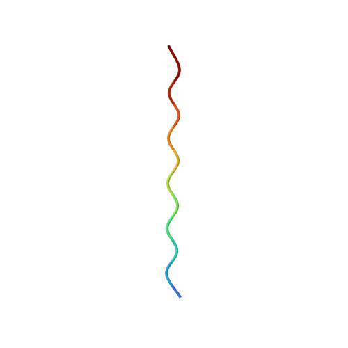
The molecular structure of the amyloid fibril has remained elusive because of the difficulty of growing well diffracting crystals. By using a sequence-designed polypeptide, we have produced crystals of an amyloid fiber. These crystals diffract to high resolution (1 A) by electron and x-ray diffraction, enabling us to determine a detailed structure for amyloid. The structure reveals that the polypeptides form fibrous crystals composed of antiparallel beta-sheets in a cross-beta arrangement, characteristic of all amyloid fibers, and allows us to determine the side-chain packing within an amyloid fiber. The antiparallel beta-sheets are zipped together by means of pi-bonding between adjacent phenylalanine rings and salt-bridges between charge pairs (glutamic acid-lysine), thus controlling and stabilizing the structure. These interactions are likely to be important in the formation and stability of other amyloid fibrils.
- Structural Medicine, Department of Haematology, University of Cambridge, Cambridge Institute for Medical Research, Hills Road, Cambridge CB2 2XY, United Kingdom.
Organizational Affiliation:


