2BEG | pdb_00002beg

3D Structure of Alzheimer's Abeta(1-42) fibrils


Experimental Data Snapshot

  • Method: SOLUTION NMR
  • Conformers Calculated: 100 
  • Conformers Submitted: 10 
  • Selection Criteria: structures with the least restraint violations 

wwPDB Validation 3D Report Full Report

Validation slider image for 2BEG

This is version 1.4 of the entry. See complete history

Literature

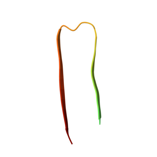
3D structure of Alzheimer's amyloid-{beta}(1-42) fibrils.

Luhrs, T.Ritter, C.Adrian, M.Riek-Loher, D.Bohrmann, B.Dobeli, H.Schubert, D.Riek, R.

(2005) Proc Natl Acad Sci U S A 102: 17342-17347

  • DOI: https://doi.org/10.1073/pnas.0506723102
  • Primary Citation Related Structures: 
    2BEG

  • PubMed Abstract: 

    Alzheimer's disease is the most fatal neurodegenerative disorder wherein the process of amyloid-beta (Abeta) amyloidogenesis appears causative. Here, we present the 3D structure of the fibrils comprising Abeta(1-42), which was obtained by using hydrogen-bonding constraints from quenched hydrogen/deuterium-exchange NMR, side-chain packing constraints from pairwise mutagenesis studies, and parallel, in-register beta-sheet arrangement from previous solid-state NMR studies. Although residues 1-17 are disordered, residues 18-42 form a beta-strand-turn-beta-strand motif that contains two intermolecular, parallel, in-register beta-sheets that are formed by residues 18-26 (beta1) and 31-42 (beta2). At least two molecules of Abeta(1-42) are required to achieve the repeating structure of a protofilament. Intermolecular side-chain contacts are formed between the odd-numbered residues of strand beta1 of the nth molecule and the even-numbered residues of strand beta2 of the (n - 1)th molecule. This interaction pattern leads to partially unpaired beta-strands at the fibrillar ends, which explains the sequence selectivity, the cooperativity, and the apparent unidirectionality of Abeta fibril growth. It also provides a structural basis for fibrillization inhibitors.


  • Organizational Affiliation
    • The Salk Institute for Biological Studies, La Jolla, CA 92037, USA.

Macromolecule Content 

  • Total Structure Weight: 22.6 kDa 
  • Atom Count: 900 
  • Modeled Residue Count: 130 
  • Deposited Residue Count: 210 
  • Unique protein chains: 1

Macromolecules

Find similar proteins by:|  3D Structure
Entity ID: 1
MoleculeChains  Sequence LengthOrganismDetailsImage
Amyloid beta A4 protein
A, B, C, D, E
42Homo sapiensMutation(s): 0 
Gene Names: APP
Membrane Entity: Yes 
UniProt & NIH Common Fund Data Resources
Find proteins for P05067 (Homo sapiens)
Explore P05067 
Go to UniProtKB:  P05067
PHAROS:  P05067
GTEx:  ENSG00000142192 
Entity Groups
Sequence Clusters30% Identity50% Identity70% Identity90% Identity95% Identity100% Identity
UniProt GroupP05067
Sequence Annotations
Expand
Reference Sequence

Experimental Data & Validation

Experimental Data

  • Method: SOLUTION NMR
  • Conformers Calculated: 100 
  • Conformers Submitted: 10 
  • Selection Criteria: structures with the least restraint violations 

Structure Validation

View Full Validation Report


Entry History 

Deposition Data

Revision History  (Full details and data files)

  • Version 1.0: 2005-11-22
    Type: Initial release
  • Version 1.1: 2008-05-01
    Changes: Version format compliance
  • Version 1.2: 2011-07-13
    Changes: Version format compliance
  • Version 1.3: 2022-03-09
    Changes: Database references, Derived calculations
  • Version 1.4: 2024-05-22
    Changes: Data collection