2BB7 | pdb_00002bb7

Mn Form Of E. coli Methionine Aminopeptidase In Complex With a quinolinyl sulfonamide inhibitor


Experimental Data Snapshot

  • Method: X-RAY DIFFRACTION
  • Resolution: 1.70 Å
  • R-Value Free: 
    0.233 (Depositor), 0.230 (DCC) 
  • R-Value Work: 
    0.187 (Depositor), 0.185 (DCC) 
  • R-Value Observed: 
    0.190 (Depositor) 

Starting Model: experimental
View more details

wwPDB Validation 3D Report Full Report

Validation slider image for 2BB7

Ligand Structure Quality Assessment 


This is version 1.3 of the entry. See complete history

Literature

Metal mediated inhibition of methionine aminopeptidase by quinolinyl sulfonamides

Huang, M.Xie, S.X.Ma, Z.Q.Hanzlik, R.P.Ye, Q.Z.

(2006) Biochem Biophys Res Commun 339: 506-513

  • DOI: https://doi.org/10.1016/j.bbrc.2005.11.042
  • Primary Citation Related Structures: 
    2BB7

  • PubMed Abstract: 

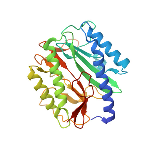
    Quinolinyl sulfonamides, such as N-(quinolin-8-yl)methanesulfonamide (10) and N-(5-chloroquinolin-8-yl)methanesulfonamide (11), were identified as potent methionine aminopeptidase (MetAP) inhibitors by high throughput screening of a diverse chemical library of small organic compounds. They showed different inhibitory potencies on Co(II)-, Ni(II)-, Fe(II)-, Mn(II)-, and Zn(II)-forms of Escherichia coli MetAP, and their inhibition is dependent on metal concentration. X-ray structures of E. coli MetAP complexed with 10 revealed that the inhibitor forms a metal complex with the residue H79 at the enzyme active site; the complex is further stabilized by an extended H-bond and metal interaction network. Analysis of the inhibition of MetAP by these inhibitors indicates that this is a typical mechanism of inhibition for many non-peptidic MetAP inhibitors and emphasizes the importance of defining in vitro conditions for identifying and evaluating MetAP inhibitors that will be capable of giving information relevant to the in vivo situation.


  • Organizational Affiliation
    • High Throughput Screening Laboratory, University of Kansas, Lawrence, KS 66045, USA.

Macromolecule Content 

  • Total Structure Weight: 29.78 kDa 
  • Atom Count: 2,246 
  • Modeled Residue Count: 258 
  • Deposited Residue Count: 264 
  • Unique protein chains: 1

Macromolecules

Find similar proteins by:|  3D Structure
Entity ID: 1
MoleculeChains  Sequence LengthOrganismDetailsImage
Methionine aminopeptidase264Escherichia coliMutation(s): 0 
Gene Names: map
EC: 3.4.11.18
UniProt
Find proteins for P0AE18 (Escherichia coli (strain K12))
Explore P0AE18 
Go to UniProtKB:  P0AE18
Entity Groups
Sequence Clusters30% Identity50% Identity70% Identity90% Identity95% Identity100% Identity
UniProt GroupP0AE18
Sequence Annotations
Expand
Reference Sequence

Experimental Data & Validation

Experimental Data

  • Method: X-RAY DIFFRACTION
  • Resolution: 1.70 Å
  • R-Value Free:  0.233 (Depositor), 0.230 (DCC) 
  • R-Value Work:  0.187 (Depositor), 0.185 (DCC) 
  • R-Value Observed: 0.190 (Depositor) 
Space Group: P 1 21 1
Unit Cell:
Length ( Å )Angle ( ˚ )
a = 38.039α = 90
b = 60.416β = 104.4
c = 50.521γ = 90
Software Package:
Software NamePurpose
SCALAdata scaling
MOLREPphasing
REFMACrefinement
PDB_EXTRACTdata extraction
MOSFLMdata reduction
CCP4data scaling

Structure Validation

View Full Validation Report


Ligand Structure Quality Assessment 


Entry History 

Deposition Data

  • Released Date: 2006-01-24 
  • Deposition Author(s): Ye, Q.Z.

Revision History  (Full details and data files)

  • Version 1.0: 2006-01-24
    Type: Initial release
  • Version 1.1: 2008-05-01
    Changes: Version format compliance
  • Version 1.2: 2011-07-13
    Changes: Version format compliance
  • Version 1.3: 2023-08-23
    Changes: Data collection, Database references, Derived calculations, Refinement description