2B9L | pdb_00002b9l

Crystal structure of prophenoloxidase activating factor-II from the beetle Holotrichia diomphalia


Experimental Data Snapshot

  • Method: X-RAY DIFFRACTION
  • Resolution: 2.00 Å
  • R-Value Free: 
    0.238 (Depositor), 0.240 (DCC) 
  • R-Value Work: 
    0.209 (Depositor), 0.210 (DCC) 
  • R-Value Observed: 
    0.209 (Depositor) 

Starting Model: experimental
View more details

wwPDB Validation   3D Report Full Report


This is version 2.2 of the entry. See complete history


Literature

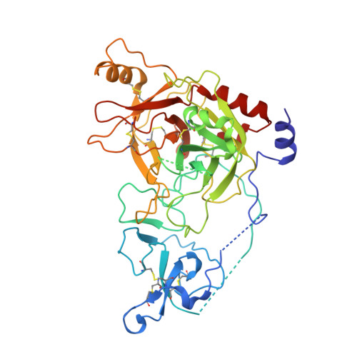
Crystal structure of a clip-domain serine protease and functional roles of the clip domains.

Piao, S.Song, Y.L.Kim, J.H.Park, S.Y.Park, J.W.Lee, B.L.Oh, B.H.Ha, N.C.

(2005) EMBO J 24: 4404-4414

  • DOI: https://doi.org/10.1038/sj.emboj.7600891
  • Primary Citation of Related Structures:  
    2B9L

  • PubMed Abstract: 

    Clip-domain serine proteases (SPs) are the essential components of extracellular signaling cascades in various biological processes, especially in embryonic development and the innate immune responses of invertebrates. They consist of a chymotrypsin-like SP domain and one or two clip domains at the N-terminus. Prophenoloxidase-activating factor (PPAF)-II, which belongs to the noncatalytic clip-domain SP family, is indispensable for the generation of the active phenoloxidase leading to melanization, a major defense mechanism of insects. Here, the crystal structure of PPAF-II reveals that the clip domain adopts a novel fold containing a central cleft, which is distinct from the structures of defensins with a similar arrangement of cysteine residues. Ensuing studies demonstrated that PPAF-II forms a homo-oligomer upon cleavage by the upstream protease and that the clip domain of PPAF-II functions as a module for binding phenoloxidase through the central cleft, while the clip domain of a catalytically active easter-type SP plays an essential role in the rapid activation of its protease domain.


  • Organizational Affiliation
    • National Research Laboratory of Defense Proteins, College of Pharmacy and Research Institute for Drug Development, Pusan National University, Jangjeon Dong, Geumjeong Gu, Busan, Korea.

Macromolecules
Find similar proteins by:  (by identity cutoff)  |  3D Structure
Entity ID: 1
MoleculeChains Sequence LengthOrganismDetailsImage
prophenoloxidase activating factor394Holotrichia diomphaliaMutation(s): 0 
UniProt
Find proteins for Q9GRW0 (Holotrichia diomphalia)
Explore Q9GRW0 
Go to UniProtKB:  Q9GRW0
Entity Groups  
Sequence Clusters30% Identity50% Identity70% Identity90% Identity95% Identity100% Identity
UniProt GroupQ9GRW0
Glycosylation
Glycosylation Sites: 1
Sequence Annotations
Expand
  • Reference Sequence
Oligosaccharides

Help

Entity ID: 2
MoleculeChains Length2D Diagram Glycosylation3D Interactions
alpha-L-fucopyranose-(1-3)-[2-acetamido-2-deoxy-beta-D-glucopyranose-(1-4)][alpha-L-fucopyranose-(1-6)]2-acetamido-2-deoxy-beta-D-glucopyranose
B
4N-Glycosylation
Glycosylation Resources
GlyTouCan:  G63564LA
GlyCosmos:  G63564LA
Small Molecules
Ligands 2 Unique
IDChains Name / Formula / InChI Key2D Diagram3D Interactions
SO4
Query on SO4

Download Ideal Coordinates CCD File 
C [auth A]
D [auth A]
E [auth A]
F [auth A]
G [auth A]
C [auth A],
D [auth A],
E [auth A],
F [auth A],
G [auth A],
H [auth A],
I [auth A]
SULFATE ION
O4 S
QAOWNCQODCNURD-UHFFFAOYSA-L
CA
Query on CA

Download Ideal Coordinates CCD File 
J [auth A]CALCIUM ION
Ca
BHPQYMZQTOCNFJ-UHFFFAOYSA-N
Experimental Data & Validation

Experimental Data

  • Method: X-RAY DIFFRACTION
  • Resolution: 2.00 Å
  • R-Value Free:  0.238 (Depositor), 0.240 (DCC) 
  • R-Value Work:  0.209 (Depositor), 0.210 (DCC) 
  • R-Value Observed: 0.209 (Depositor) 
Space Group: C 1 2 1
Unit Cell:
Length ( Å )Angle ( ˚ )
a = 106.78α = 90
b = 76.587β = 113.45
c = 69.833γ = 90
Software Package:
Software NamePurpose
CNSrefinement
HKL-2000data reduction
SCALEPACKdata scaling
MOLREPphasing

Structure Validation

View Full Validation Report


Entry History 

Deposition Data

Revision History  (Full details and data files)

  • Version 1.0: 2006-01-03
    Type: Initial release
  • Version 1.1: 2008-05-01
    Changes: Version format compliance
  • Version 1.2: 2011-07-13
    Changes: Non-polymer description, Version format compliance
  • Version 2.0: 2020-07-29
    Type: Remediation
    Reason: Carbohydrate remediation
    Changes: Atomic model, Data collection, Database references, Derived calculations, Structure summary
  • Version 2.1: 2023-08-23
    Changes: Data collection, Database references, Refinement description, Structure summary
  • Version 2.2: 2024-10-30
    Changes: Structure summary