2ASP

Structure of Rabbit Actin In Complex With Reidispongiolide C


Experimental Data Snapshot

  • Method: X-RAY DIFFRACTION
  • Resolution: 1.64 Å
  • R-Value Free: 0.193 
  • R-Value Work: 0.163 
  • R-Value Observed: 0.164 

wwPDB Validation   3D Report Full Report


Ligand Structure Quality Assessment 


This is version 1.3 of the entry. See complete history


Literature

Structures of microfilament destabilizing toxins bound to actin provide insight into toxin design and activity

Allingham, J.S.Zampella, A.D'Auria, M.V.Rayment, I.

(2005) Proc Natl Acad Sci U S A 102: 14527-14532

  • DOI: https://doi.org/10.1073/pnas.0502089102
  • Primary Citation of Related Structures:  
    2ASM, 2ASO, 2ASP

  • PubMed Abstract: 

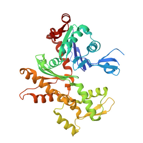
    Marine macrolides that disrupt the actin cytoskeleton are promising candidates for cancer treatment. Here, we present the actin-bound x-ray crystal structures of reidispongiolide A and C and sphinxolide B, three marine macrolides found among a recently discovered family of cytotoxic compounds. Their structures allow unequivocal assignment of the absolute configuration for each compound. A comparison of their actin-binding site to macrolides found in the trisoxazole family, as well as the divalent macrolide, swinholide A, reveals the existence of a common binding surface for a defined segment of their macrocyclic ring. This surface is located on a hydrophobic patch adjacent to the cleft separating domains 1 and 3 at the barbed-end of actin. The large area surrounding this surface accommodates a wide variety of conformations and designs observed in the macrocyclic component of barbed-end-targeting macrolides. Conversely, the binding pocket for the macrolide tail, located within the cleft itself, shows very limited variation. Functional characterization of these macrolides by using in vitro actin filament severing and polymerization assays demonstrate the necessity of the N-methyl-vinylformamide moiety at the terminus of the macrolide tail for toxin potency. These analyses also show the importance of stable interactions between the macrocyclic ring and the hydrophobic patch on actin for modifying filament structure and how this stability can be compromised by subtle changes in macrolactone ring composition. By identifying the essential components of these complex natural products that underlie their high actin affinity, we have established a framework for designing new therapeutic agents.


  • Organizational Affiliation

    Department of Biochemistry, University of Wisconsin, Madison, WI 53706-1544, USA.


Macromolecules
Find similar proteins by:  (by identity cutoff)  |  3D Structure
Entity ID: 1
MoleculeChains Sequence LengthOrganismDetailsImage
Actin, alpha skeletal muscle375Oryctolagus cuniculusMutation(s): 0 
UniProt
Find proteins for P68135 (Oryctolagus cuniculus)
Explore P68135 
Go to UniProtKB:  P68135
Entity Groups  
Sequence Clusters30% Identity50% Identity70% Identity90% Identity95% Identity100% Identity
UniProt GroupP68135
Sequence Annotations
Expand
  • Reference Sequence
Small Molecules
Ligands 4 Unique
IDChains Name / Formula / InChI Key2D Diagram3D Interactions
RGC
Query on RGC

Download Ideal Coordinates CCD File 
E [auth A]REIDISPONGIOLIDE C
C52 H84 O14
QTLSHCJDXCYLJK-VRWAWWQJSA-N
ATP
Query on ATP

Download Ideal Coordinates CCD File 
D [auth A]ADENOSINE-5'-TRIPHOSPHATE
C10 H16 N5 O13 P3
ZKHQWZAMYRWXGA-KQYNXXCUSA-N
EDO
Query on EDO

Download Ideal Coordinates CCD File 
F [auth A],
G [auth A],
H [auth A],
I [auth A],
J [auth A]
1,2-ETHANEDIOL
C2 H6 O2
LYCAIKOWRPUZTN-UHFFFAOYSA-N
CA
Query on CA

Download Ideal Coordinates CCD File 
B [auth A],
C [auth A]
CALCIUM ION
Ca
BHPQYMZQTOCNFJ-UHFFFAOYSA-N
Experimental Data & Validation

Experimental Data

  • Method: X-RAY DIFFRACTION
  • Resolution: 1.64 Å
  • R-Value Free: 0.193 
  • R-Value Work: 0.163 
  • R-Value Observed: 0.164 
  • Space Group: C 1 2 1
Unit Cell:
Length ( Å )Angle ( ˚ )
a = 170.402α = 90
b = 54.689β = 95.81
c = 40.872γ = 90
Software Package:
Software NamePurpose
REFMACrefinement
HKL-2000data reduction
SCALEPACKdata scaling
MOLREPphasing

Structure Validation

View Full Validation Report


Ligand Structure Quality Assessment 


Entry History 

Deposition Data

Revision History  (Full details and data files)

  • Version 1.0: 2005-10-11
    Type: Initial release
  • Version 1.1: 2008-04-30
    Changes: Version format compliance
  • Version 1.2: 2011-07-13
    Changes: Version format compliance
  • Version 1.3: 2023-10-25
    Changes: Data collection, Database references, Derived calculations, Refinement description, Structure summary