2AJY

Crystal Structure of Cocaine catalytic Antibody 7A1 Fab' in Complex with ecgonine methyl ester and benzoic acid


Experimental Data Snapshot

  • Method: X-RAY DIFFRACTION
  • Resolution: 2.10 Å
  • R-Value Free: 0.246 
  • R-Value Work: 0.225 

wwPDB Validation   3D Report Full Report


Ligand Structure Quality Assessment 


This is version 1.4 of the entry. See complete history


Literature

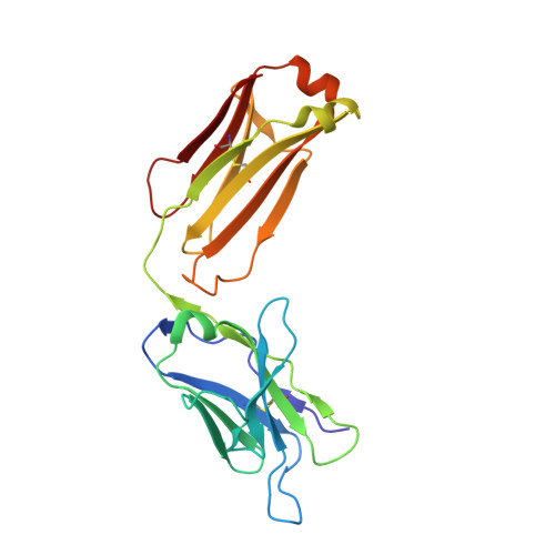
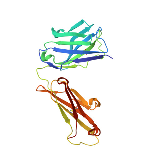
Complete reaction cycle of a cocaine catalytic antibody at atomic resolution.

Zhu, X.Dickerson, T.J.Rogers, C.J.Kaufmann, G.F.Mee, J.M.McKenzie, K.M.Janda, K.D.Wilson, I.A.

(2006) Structure 14: 205-216

  • DOI: https://doi.org/10.1016/j.str.2005.10.014
  • Primary Citation of Related Structures:  
    2AJS, 2AJU, 2AJV, 2AJX, 2AJY, 2AJZ, 2AK1

  • PubMed Abstract: 

    Antibody 7A1 hydrolyzes cocaine to produce nonpsychoactive metabolites ecgonine methyl ester and benzoic acid. Crystal structures of 7A1 Fab' and six complexes with substrate cocaine, the transition state analog, products ecgonine methyl ester and benzoic acid together and individually, as well as heptaethylene glycol have been analyzed at 1.5-2.3 angstroms resolution. Here, we present snapshots of the complete cycle of the cocaine hydrolytic reaction at atomic resolution. Significant structural rearrangements occur along the reaction pathway, but they are generally limited to the binding site, including the ligands themselves. Several interacting side chains either change their rotamers or alter their mobility to accommodate the different reaction steps. CDR loop movements (up to 2.3 angstroms) and substantial side chain rearrangements (up to 9 angstroms) alter the shape and size (approximately 320-500 angstroms3) of the antibody active site from "open" to "closed" to "open" for the substrate, transition state, and product states, respectively.


  • Organizational Affiliation

    Department of Molecular Biology, The Scripps Research Institute, 10550 North Torrey Pines Road, La Jolla, California 92037, USA.


Macromolecules
Find similar proteins by:  (by identity cutoff)  |  3D Structure
Entity ID: 1
MoleculeChains Sequence LengthOrganismDetailsImage
Antibody 7A1 Fab'A [auth L]216Mus musculusMutation(s): 0 
UniProt
Find proteins for P01837 (Mus musculus)
Explore P01837 
Go to UniProtKB:  P01837
Entity Groups  
Sequence Clusters30% Identity50% Identity70% Identity90% Identity95% Identity100% Identity
UniProt GroupP01837
Sequence Annotations
Expand
  • Reference Sequence
Find similar proteins by:  (by identity cutoff)  |  3D Structure
Entity ID: 2
MoleculeChains Sequence LengthOrganismDetailsImage
Antibody 7A1 Fab'B [auth H]219Mus musculusMutation(s): 0 
UniProt
Find proteins for Q5M842 (Rattus norvegicus)
Explore Q5M842 
Go to UniProtKB:  Q5M842
Entity Groups  
Sequence Clusters30% Identity50% Identity70% Identity90% Identity95% Identity100% Identity
UniProt GroupQ5M842
Sequence Annotations
Expand
  • Reference Sequence
Experimental Data & Validation

Experimental Data

  • Method: X-RAY DIFFRACTION
  • Resolution: 2.10 Å
  • R-Value Free: 0.246 
  • R-Value Work: 0.225 
  • Space Group: C 2 2 21
Unit Cell:
Length ( Å )Angle ( ˚ )
a = 83.569α = 90
b = 89.802β = 90
c = 154.446γ = 90
Software Package:
Software NamePurpose
DENZOdata reduction
SCALEPACKdata scaling
CNSrefinement
PDB_EXTRACTdata extraction
HKL-2000data reduction
HKL-2000data scaling
AMoREphasing

Structure Validation

View Full Validation Report


Ligand Structure Quality Assessment 


Entry History 

Deposition Data

Revision History  (Full details and data files)

  • Version 1.0: 2006-02-14
    Type: Initial release
  • Version 1.1: 2008-04-30
    Changes: Version format compliance
  • Version 1.2: 2011-07-13
    Changes: Version format compliance
  • Version 1.3: 2017-10-11
    Changes: Refinement description
  • Version 1.4: 2023-08-23
    Changes: Data collection, Database references, Derived calculations, Refinement description