2AEI | pdb_00002aei

Crystal structure of a ternary complex of factor VIIa/tissue factor and 2-[[6-[3-(aminoiminomethyl)phenoxy]-3,5-difluro-4-[(1-methyl-3-phenylpropyl)amino]-2-pyridinyl]oxy]-benzoic acid


Experimental Data Snapshot

  • Method: X-RAY DIFFRACTION
  • Resolution: 2.52 Å
  • R-Value Free: 
    0.297 (Depositor), 0.288 (DCC) 
  • R-Value Work: 
    0.199 (Depositor), 0.208 (DCC) 
  • R-Value Observed: 
    0.204 (Depositor) 

Starting Model: experimental
View more details

wwPDB Validation 3D Report Full Report

Validation slider image for 2AEI

Ligand Structure Quality Assessment 


This is version 1.4 of the entry. See complete history

Literature

The discovery of fluoropyridine-based inhibitors of the Factor VIIa/TF complex.

Kohrt, J.T.Filipski, K.J.Cody, W.L.Cai, C.Dudley, D.A.Van Huis, C.A.Willardsen, J.A.Rapundalo, S.T.Saiya-Cork, K.Leadley, R.J.Narasimhan, L.Zhang, E.Whitlow, M.Adler, M.McLean, K.Chou, Y.L.McKnight, C.Arnaiz, D.O.Shaw, K.J.Light, D.R.Edmunds, J.J.

(2005) Bioorg Med Chem Lett 15: 4752-4756

  • DOI: https://doi.org/10.1016/j.bmcl.2005.07.059
  • Primary Citation Related Structures: 
    2AEI

  • PubMed Abstract: 

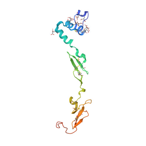
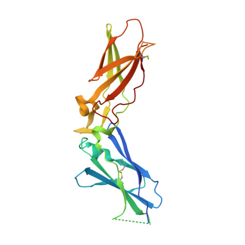
    The activated Factor VII/tissue factor complex (FVIIa/TF) plays a key role in the formation of blood clots. Inhibition of this complex may lead to new antithrombotic drugs. An X-ray crystal structure of a fluoropyridine-based FVIIa/TF inhibitor bound in the active site of the enzyme complex suggested that incorporation of substitution at the 5-position of the hydroxybenzoic acid side chain could lead to the formation of more potent inhibitors through interactions with the S1'/S2' pocket.


  • Organizational Affiliation
    • Pfizer Global Research and Development, Michigan Laboratories, 2800 Plymouth Road, Ann Arbor, MI 48105, USA. jeffrey.kohrt@pfizer.com

Macromolecule Content 

  • Total Structure Weight: 70.4 kDa 
  • Atom Count: 4,812 
  • Modeled Residue Count: 591 
  • Deposited Residue Count: 617 
  • Unique protein chains: 3

Macromolecules

Find similar proteins by:|  3D Structure
Entity ID: 1
MoleculeChains  Sequence LengthOrganismDetailsImage
Coagulation factor VIIA [auth L]152Homo sapiensMutation(s): 10 
Gene Names: F7
EC: 3.4.21.21
UniProt & NIH Common Fund Data Resources
Find proteins for P08709 (Homo sapiens)
Explore P08709 
Go to UniProtKB:  P08709
PHAROS:  P08709
GTEx:  ENSG00000057593 
Entity Groups
Sequence Clusters30% Identity50% Identity70% Identity90% Identity95% Identity100% Identity
UniProt GroupP08709
Sequence Annotations
Expand
Reference Sequence
Find similar proteins by:|  3D Structure
Entity ID: 2
MoleculeChains  Sequence LengthOrganismDetailsImage
Coagulation factor VIIB [auth H]254Homo sapiensMutation(s): 0 
Gene Names: F7
EC: 3.4.21.21
UniProt & NIH Common Fund Data Resources
Find proteins for P08709 (Homo sapiens)
Explore P08709 
Go to UniProtKB:  P08709
PHAROS:  P08709
GTEx:  ENSG00000057593 
Entity Groups
Sequence Clusters30% Identity50% Identity70% Identity90% Identity95% Identity100% Identity
UniProt GroupP08709
Sequence Annotations
Expand
Reference Sequence
Find similar proteins by:|  3D Structure
Entity ID: 3
MoleculeChains  Sequence LengthOrganismDetailsImage
Tissue factorC [auth T]211Homo sapiensMutation(s): 0 
Gene Names: F3
UniProt & NIH Common Fund Data Resources
Find proteins for P13726 (Homo sapiens)
Explore P13726 
Go to UniProtKB:  P13726
PHAROS:  P13726
GTEx:  ENSG00000117525 
Entity Groups
Sequence Clusters30% Identity50% Identity70% Identity90% Identity95% Identity100% Identity
UniProt GroupP13726
Sequence Annotations
Expand
Reference Sequence

Small Molecules

Ligands 3 Unique
IDChains Name / Formula / InChI Key2D Diagram3D Interactions
03R

Query on 03R


Download:Ideal Coordinates CCD File
M [auth H]2-({6-{3-[AMINO(IMINO)METHYL]PHENOXY}-3,5-DIFLUORO-4-[(1-METHYL-3-PHENYLPROPYL)AMINO]-2-PYRIDINYL}OXY)BENZOIC ACID
C29 H26 F2 N4 O4
BUZNVJZUYOBGFM-KRWDZBQOSA-N
CAC

Query on CAC


Download:Ideal Coordinates CCD File
L [auth H]CACODYLATE ION
C2 H6 As O2
OGGXGZAMXPVRFZ-UHFFFAOYSA-M
CA

Query on CA


Download:Ideal Coordinates CCD File
D [auth L]
E [auth L]
F [auth L]
G [auth L]
H [auth L]
D [auth L],
E [auth L],
F [auth L],
G [auth L],
H [auth L],
I [auth L],
J [auth L],
K [auth H]
CALCIUM ION
Ca
BHPQYMZQTOCNFJ-UHFFFAOYSA-N
Modified Residues  1 Unique
IDChains TypeFormula2D DiagramParent
CGU
Query on CGU
A [auth L]L-PEPTIDE LINKINGC6 H9 N O6GLU
Binding Affinity Annotations 
IDSourceBinding Affinity
03R BindingDB:  2AEI IC50: 86 (nM) from 1 assay(s)

Experimental Data & Validation

Experimental Data

  • Method: X-RAY DIFFRACTION
  • Resolution: 2.52 Å
  • R-Value Free:  0.297 (Depositor), 0.288 (DCC) 
  • R-Value Work:  0.199 (Depositor), 0.208 (DCC) 
  • R-Value Observed: 0.204 (Depositor) 
Space Group: P 21 21 21
Unit Cell:
Length ( Å )Angle ( ˚ )
a = 70.215α = 90
b = 80.496β = 90
c = 125.936γ = 90
Software Package:
Software NamePurpose
MOSFLMdata reduction
SCALAdata scaling
MERLOTphasing
X-PLORrefinement
DENZOdata reduction
SCALEPACKdata scaling

Structure Validation

View Full Validation Report


Ligand Structure Quality Assessment 


Entry History 

Deposition Data

Revision History  (Full details and data files)

  • Version 1.0: 2006-08-01
    Type: Initial release
  • Version 1.1: 2008-04-30
    Changes: Version format compliance
  • Version 1.2: 2011-07-13
    Changes: Non-polymer description, Version format compliance
  • Version 1.3: 2023-08-23
    Changes: Data collection, Database references, Derived calculations, Refinement description
  • Version 1.4: 2023-11-15
    Changes: Data collection