1ZUI

Structural Basis for Shikimate-binding Specificity of Helicobacter pylori Shikimate Kinase


Experimental Data Snapshot

  • Method: X-RAY DIFFRACTION
  • Resolution: 2.30 Å
  • R-Value Free: 0.280 
  • R-Value Work: 0.203 
  • R-Value Observed: 0.207 

wwPDB Validation   3D Report Full Report


Ligand Structure Quality Assessment 


This is version 1.4 of the entry. See complete history


Literature

Structural basis for shikimate-binding specificity of Helicobacter pylori shikimate kinase

Cheng, W.C.Chang, Y.N.Wang, W.C.

(2005) J Bacteriol 187: 8156-8163

  • DOI: https://doi.org/10.1128/JB.187.23.8156-8163.2005
  • Primary Citation of Related Structures:  
    1ZUH, 1ZUI

  • PubMed Abstract: 

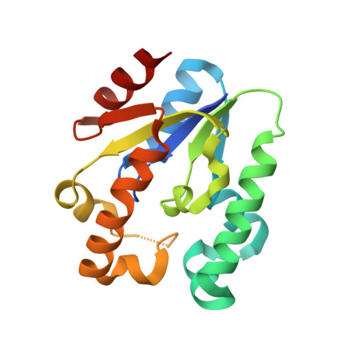
    Shikimate kinase (EC 2.7.1.71) catalyzes the specific phosphorylation of the 3-hydroxyl group of shikimic acid in the presence of ATP. As the fifth key step in the shikimate pathway for aromatic amino acid biosynthesis in bacteria, fungi, and plants, but not mammals, shikimate kinase represents an attractive target for the development of new antimicrobial agents, herbicides, and antiparasitic agents. Here, we report the 1.8-Angstroms crystal structure of Helicobacter pylori shikimate kinase (HpSK). The crystal structure shows a three-layer alpha/beta fold consisting of a central sheet of five parallel beta-strands flanked by seven alpha-helices. An HpSK-shikimate-PO(4) complex was also determined and refined to 2.3 Angstroms, revealing induced-fit movement from an open to a closed form on substrate binding. Shikimate is located above a short 3(10) helix formed by a strictly conserved motif (GGGXV) after beta(3). Moreover, several highly conserved charged residues including Asp33 (in a conserved DT/SD motif), Arg57, and Arg132 (interacting with shikimate) are identified, guiding the development of novel inhibitors of shikimate kinase.


  • Organizational Affiliation

    Institute of Molecular and Cellular Biology and Department of Life Sciences, National Tsing Hua University, Hsinchu, Taiwan.


Macromolecules
Find similar proteins by:  (by identity cutoff)  |  3D Structure
Entity ID: 1
MoleculeChains Sequence LengthOrganismDetailsImage
Shikimate kinase168Helicobacter pylori 26695Mutation(s): 0 
Gene Names: AroK
EC: 2.7.1.71
UniProt
Find proteins for P56073 (Helicobacter pylori (strain ATCC 700392 / 26695))
Explore P56073 
Go to UniProtKB:  P56073
Entity Groups  
Sequence Clusters30% Identity50% Identity70% Identity90% Identity95% Identity100% Identity
UniProt GroupP56073
Sequence Annotations
Expand
  • Reference Sequence
Small Molecules
Ligands 2 Unique
IDChains Name / Formula / InChI Key2D Diagram3D Interactions
SKM
Query on SKM

Download Ideal Coordinates CCD File 
C [auth A](3R,4S,5R)-3,4,5-TRIHYDROXYCYCLOHEX-1-ENE-1-CARBOXYLIC ACID
C7 H10 O5
JXOHGGNKMLTUBP-HSUXUTPPSA-N
PO4
Query on PO4

Download Ideal Coordinates CCD File 
B [auth A]PHOSPHATE ION
O4 P
NBIIXXVUZAFLBC-UHFFFAOYSA-K
Experimental Data & Validation

Experimental Data

  • Method: X-RAY DIFFRACTION
  • Resolution: 2.30 Å
  • R-Value Free: 0.280 
  • R-Value Work: 0.203 
  • R-Value Observed: 0.207 
  • Space Group: P 61
Unit Cell:
Length ( Å )Angle ( ˚ )
a = 97.286α = 90
b = 97.286β = 90
c = 46.909γ = 120
Software Package:
Software NamePurpose
DENZOdata reduction
SCALEPACKdata scaling
MOLREPphasing
REFMACrefinement
PDB_EXTRACTdata extraction

Structure Validation

View Full Validation Report


Ligand Structure Quality Assessment 


Entry History 

Deposition Data

Revision History  (Full details and data files)

  • Version 1.0: 2006-05-31
    Type: Initial release
  • Version 1.1: 2008-04-30
    Changes: Version format compliance
  • Version 1.2: 2011-07-13
    Changes: Source and taxonomy, Version format compliance
  • Version 1.3: 2017-10-11
    Changes: Refinement description
  • Version 1.4: 2024-03-13
    Changes: Data collection, Database references, Derived calculations