Catalytic cycling in beta-phosphoglucomutase: a kinetic and structural analysis
Zhang, G., Dai, J., Wang, L., Dunaway-Mariano, D., Tremblay, L.W., Allen, K.N.(2005) Biochemistry 44: 9404-9416
- PubMed: 15996095 Search on PubMed
- DOI: https://doi.org/10.1021/bi050558p
- Primary Citation Related Structures:
1ZOL - PubMed Abstract:
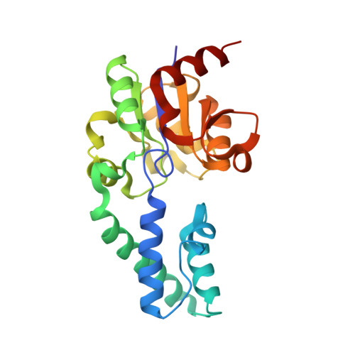
Lactococcus lactis beta-phosphoglucomutase (beta-PGM) catalyzes the interconversion of beta-d-glucose 1-phosphate (beta-G1P) and beta-d-glucose 6-phosphate (G6P), forming beta-d-glucose 1,6-(bis)phosphate (beta-G16P) as an intermediate. Beta-PGM conserves the core domain catalytic scaffold of the phosphatase branch of the HAD (haloalkanoic acid dehalogenase) enzyme superfamily, yet it has evolved to function as a mutase rather than as a phosphatase. This work was carried out to identify the structural basis underlying this diversification of function. In this paper, we examine beta-PGM activation by the Mg(2+) cofactor, beta-PGM activation by Asp8 phosphorylation, and the role of cap domain closure in substrate discrimination. First, the 1.90 A resolution X-ray crystal structure of the Mg(2+)-beta-PGM complex is examined in the context of previously reported structures of the Mg(2+)-alpha-d-galactose-1-phosphate-beta-PGM, Mg(2+)-phospho-beta-PGM, and Mg(2+)-beta-glucose-6-phosphate-1-phosphorane-beta-PGM complexes to identify conformational changes that occur during catalytic turnover. The essential role of Asp8 in nucleophilic catalysis was confirmed by demonstrating that the D8A and D8E mutants are devoid of catalytic activity. Comparison of the ligands to Mg(2+) in the different complexes shows that a single Mg(2+) coordination site must alternatively accommodate water, phosphate, and the phosphorane intermediate during catalytic turnover. Limited involvement of the HAD family metal-binding loop in Mg(2+) anchoring in beta-PGM is consistent with the relatively loose binding indicated by the large K(m) for Mg(2+) activation (270 +/- 20 microM) and with the retention of activity found in the E169A/D170A double loop mutant. Comparison of the relative positions of cap and core domains in the different complexes indicated that interaction of cap domain Arg49 with the "nontransferring" phosphoryl group of the substrate ligand might stabilize the cap-closed conformation, as required for active site desolvation and alignment of Asp10 for acid-base catalysis. Kinetic analyses of the specificity of beta-PGM toward phosphoryl group donors and the specificity of phospho-beta-PGM toward phosphoryl group acceptors were carried out. The results support a substrate induced-fit mechanism of beta-PGM catalysis, which allows phosphomutase activity to dominate over the intrinsic phosphatase activity. Last, we present evidence that the autophosphorylation of beta-PGM by the substrate beta-G1P accounts for the origin of phospho-beta-PGM in the cell.
- Department of Chemistry, University of New Mexico, Albuquerque, New Mexico 87131-0001, USA.
Organizational Affiliation: