1WU1

Factor Xa in complex with the inhibitor 4-[(5-chloroindol-2-yl)sulfonyl]-2-(2-methylpropyl)-1-[[5-(pyridin-4-yl) pyrimidin-2-yl]carbonyl]piperazine


Experimental Data Snapshot

  • Method: X-RAY DIFFRACTION
  • Resolution: 2.30 Å
  • R-Value Free: 0.235 
  • R-Value Work: 0.189 
  • R-Value Observed: 0.191 

wwPDB Validation   3D Report Full Report


This is version 1.3 of the entry. See complete history


Literature

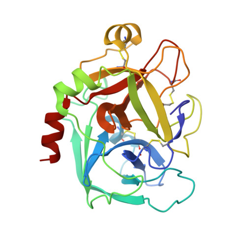
Design, synthesis, and biological activity of non-basic compounds as factor Xa inhibitors: SAR study of S1 and aryl binding sites

Komoriya, S.Haginoya, N.Kobayashi, S.Nagata, T.Mochizuki, A.Suzuki, M.Yoshino, T.Horino, H.Nagahara, T.Suzuki, M.Isobe, Y.Furugoori, T.

(2005) Bioorg Med Chem 13: 3927-3954

  • DOI: 10.1016/j.bmc.2005.04.006
  • Primary Citation of Related Structures:  
    1WU1, 2D1J

  • PubMed Abstract: 
  • Compound 7 was identified as the active metabolite of 6 by HPLC and mass spectral analysis. Modification of lead compound 7 by transformation of its N-oxide 6-6 biaryl ring system and fused aromatics produced a series of non-basic fXa inhibitors with excellent potency in anti-fXa and anticoagulant assays ...

    Compound 7 was identified as the active metabolite of 6 by HPLC and mass spectral analysis. Modification of lead compound 7 by transformation of its N-oxide 6-6 biaryl ring system and fused aromatics produced a series of non-basic fXa inhibitors with excellent potency in anti-fXa and anticoagulant assays. The optimized compounds 73b and 75b showed sub to one digit micromolar anticoagulant activity (PTCT2). Particularly, anti-fXa activity was detected in plasma of rats orally administered with 1mg/kg of compound 75b.


    Related Citations: 
    • Structural basis for chemical inhibition of human blood coagulation factor Xa
      Kamata, K., Kawamoto, H., Honma, T., Iwama, T., Kim, S.H.
      (1998) Proc Natl Acad Sci U S A 95: 6630
    • X-ray structure of active site-inhibited clotting factor Xa. Implications for drug design and substrate recognition
      Brandstetter, H., Kuhne, A., Bode, W., Huber, R., von der Saal, W., Wirthensohn, K., Engh, R.A.
      (1996) J Biol Chem 271: 29988
    • Structure of human des(1-45) factor Xa at 2.2 A resolution
      Padmanabhan, K., Padmanabhan, K.P., Tulinsky, A., Park, C.H., Bode, W., Huber, R., Blankenship, D.T., Cardin, A.D., Kisiel, W.
      (1993) J Mol Biol 232: 947
    • Synthesis and Conformational Analysis of a Non-Amidine Factor Xa Inhibitor That Incorporates 5-Methyl-4,5,6,7-tetrahydrothiazolo[5,4-c]pyridine as S4 Binding Element
      Haginoya, N., Kobayashi, S., Komoriya, S., Yoshino, T., Suzuki, M., Shimada, T., Watanabe, K., Hirokawa, Y., Furugori, T., Nagahara, T.
      (2004) J Med Chem 47: 5167

    Organizational Affiliation

    Tokyo R&D Center, Daiichi Pharmaceutical Co. Ltd, 16-13 Kita-Kasai 1-Chome, Edogawa-ku, Tokyo 134-8630, Japan. komor0ni@daiichipharm.co.jp


Macromolecules
Find similar proteins by:  (by identity cutoff)  |  3D Structure
Entity ID: 1
MoleculeChainsSequence LengthOrganismDetailsImage
Coagulation factor X, heavy chainA233Homo sapiensMutation(s): 0 
Gene Names: F10
EC: 3.4.21.6
UniProt & NIH Common Fund Data Resources
Find proteins for P00742 (Homo sapiens)
Explore P00742 
Go to UniProtKB:  P00742
PHAROS:  P00742
Entity Groups  
Sequence Clusters30% Identity50% Identity70% Identity90% Identity95% Identity100% Identity
UniProt GroupP00742
Protein Feature View
Expand
  • Reference Sequence
Find similar proteins by:  (by identity cutoff)  |  3D Structure
Entity ID: 2
MoleculeChainsSequence LengthOrganismDetailsImage
Coagulation factor X, light chainB95Homo sapiensMutation(s): 0 
Gene Names: F10
EC: 3.4.21.6
UniProt & NIH Common Fund Data Resources
Find proteins for P00742 (Homo sapiens)
Explore P00742 
Go to UniProtKB:  P00742
PHAROS:  P00742
Entity Groups  
Sequence Clusters30% Identity50% Identity70% Identity90% Identity95% Identity100% Identity
UniProt GroupP00742
Protein Feature View
Expand
  • Reference Sequence
Small Molecules
Ligands 2 Unique
IDChainsName / Formula / InChI Key2D Diagram3D Interactions
D91
Query on D91

Download Ideal Coordinates CCD File 
E [auth A]5-CHLORO-2-({3-ISOBUTYL-4-[(5-PYRIDIN-4-YLPYRIMIDIN-2-YL)CARBONYL]PIPERAZIN-1-YL}SULFONYL)-1H-INDOLE
C26 H27 Cl N6 O3 S
JUUFKVAFKAKWRV-JOCHJYFZSA-N
 Ligand Interaction
CA
Query on CA

Download Ideal Coordinates CCD File 
C [auth A],
D [auth A]
CALCIUM ION
Ca
BHPQYMZQTOCNFJ-UHFFFAOYSA-N
 Ligand Interaction
Modified Residues  1 Unique
IDChainsTypeFormula2D DiagramParent
BHD
Query on BHD
B L-PEPTIDE LINKINGC4 H7 N O5ASP
Binding Affinity Annotations 
IDSourceBinding Affinity
D91 PDBBind:  1WU1 IC50: 18 (nM) from 1 assay(s)
Binding MOAD:  1WU1 IC50: 18 (nM) from 1 assay(s)
Experimental Data & Validation

Experimental Data

  • Method: X-RAY DIFFRACTION
  • Resolution: 2.30 Å
  • R-Value Free: 0.235 
  • R-Value Work: 0.189 
  • R-Value Observed: 0.191 
  • Space Group: P 21 21 21
Unit Cell:
Length ( Å )Angle ( ˚ )
a = 56.569α = 90
b = 72.487β = 90
c = 79.02γ = 90
Software Package:
Software NamePurpose
d*TREKdata scaling
d*TREKdata reduction
REFMACrefinement

Structure Validation

View Full Validation Report


Entry History 

Deposition Data

  • Released Date: 2005-11-29 
  • Deposition Author(s): Suzuki, M.

Revision History  (Full details and data files)

  • Version 1.0: 2005-11-29
    Type: Initial release
  • Version 1.1: 2008-04-01
    Changes: Version format compliance
  • Version 1.2: 2011-07-13
    Changes: Version format compliance
  • Version 1.3: 2012-06-20
    Changes: Non-polymer description