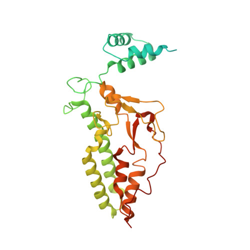
1V0D

Crystal Structure of Caspase-activated DNase (CAD)


Experimental Data Snapshot

  • Method: X-RAY DIFFRACTION
  • Resolution: 2.60 Å
  • R-Value Free: 0.246 
  • R-Value Work: 0.218 
  • R-Value Observed: 0.218 

wwPDB Validation   3D Report Full Report


This is version 1.2 of the entry. See complete history


Literature

Structural Mechanism for Inactivation and Activation of Cad/Dff40 in the Apoptotic Pathway

Woo, E.-J.Kim, Y.-G.Kim, M.-S.Han, W.-D.Shin, S.Robinson, H.Park, S.-Y.Oh, B.-H.

(2004) Mol Cell 14: 531

  • DOI: https://doi.org/10.1016/s1097-2765(04)00258-8
  • Primary Citation of Related Structures:  
    1V0D

  • PubMed Abstract: 
  • CAD/DFF40 is responsible for the degradation of chromosomal DNA into nucleosomal fragments and subsequent chromatin condensation during apoptosis. It exists as an inactive complex with its inhibitor ICAD/DFF45 in proliferating cells but becomes activated upon cleavage of ICAD/DFF45 into three domains by caspases in dying cells ...

    CAD/DFF40 is responsible for the degradation of chromosomal DNA into nucleosomal fragments and subsequent chromatin condensation during apoptosis. It exists as an inactive complex with its inhibitor ICAD/DFF45 in proliferating cells but becomes activated upon cleavage of ICAD/DFF45 into three domains by caspases in dying cells. The molecular mechanism underlying the control and activation of CAD/DFF40 was unknown. Here, the crystal structure of activated CAD/DFF40 reveals that it is a pair of molecular scissors with a deep active-site crevice that appears ideal for distinguishing internucleosomal DNA from nucleosomal DNA. Ensuing studies show that ICAD/DFF45 sequesters the nonfunctional CAD/DFF40 monomer and is also able to disassemble the functional CAD/DFF40 dimer. This capacity requires the involvement of the middle domain of ICAD/DFF45, which by itself cannot remain bound to CAD/DFF40 due to low binding affinity for the enzyme. Thus, the consequence of the caspase-cleavage of ICAD/DFF45 is a self-assembly of CAD/DFF40 into the active dimer.


    Organizational Affiliation

    Division of Molecular and Life Sciences, Department of Life Sciences and Center for Biomolecular Recognition, Pohang University of Science and Technology, Pohang, Kyungbuk 790-784, South Korea.


Macromolecules
Find similar proteins by:  (by identity cutoff)  |  3D Structure
Entity ID: 1
MoleculeChainsSequence LengthOrganismDetailsImage
DNA FRAGMENTATION FACTOR 40 KDA SUBUNIT329Mus musculusMutation(s): 0 
Gene Names: DffbCad
EC: 3
UniProt & NIH Common Fund Data Resources
Find proteins for O54788 (Mus musculus)
Explore O54788 
Go to UniProtKB:  O54788
IMPC:  MGI:1196287
Entity Groups  
Sequence Clusters30% Identity50% Identity70% Identity90% Identity95% Identity100% Identity
UniProt GroupO54788
Protein Feature View
Expand
  • Reference Sequence
Small Molecules
Ligands 3 Unique
IDChainsName / Formula / InChI Key2D Diagram3D Interactions
PB
Query on PB

Download Ideal Coordinates CCD File 
D [auth A]LEAD (II) ION
Pb
RVPVRDXYQKGNMQ-UHFFFAOYSA-N
 Ligand Interaction
ZN
Query on ZN

Download Ideal Coordinates CCD File 
B [auth A]ZINC ION
Zn
PTFCDOFLOPIGGS-UHFFFAOYSA-N
 Ligand Interaction
MG
Query on MG

Download Ideal Coordinates CCD File 
C [auth A]MAGNESIUM ION
Mg
JLVVSXFLKOJNIY-UHFFFAOYSA-N
 Ligand Interaction
Experimental Data & Validation

Experimental Data

  • Method: X-RAY DIFFRACTION
  • Resolution: 2.60 Å
  • R-Value Free: 0.246 
  • R-Value Work: 0.218 
  • R-Value Observed: 0.218 
  • Space Group: P 64
Unit Cell:
Length ( Å )Angle ( ˚ )
a = 110.189α = 90
b = 110.189β = 90
c = 73.982γ = 120
Software Package:
Software NamePurpose
CNSrefinement
SCALEPACKdata scaling

Structure Validation

View Full Validation Report


Entry History 

Deposition Data

Revision History  (Full details and data files)

  • Version 1.0: 2004-05-21
    Type: Initial release
  • Version 1.1: 2011-05-07
    Changes: Version format compliance
  • Version 1.2: 2011-07-13
    Changes: Version format compliance