1UVQ

Crystal structure of HLA-DQ0602 in complex with a hypocretin peptide


Experimental Data Snapshot

  • Method: X-RAY DIFFRACTION
  • Resolution: 1.80 Å
  • R-Value Free: 0.205 
  • R-Value Work: 0.189 
  • R-Value Observed: 0.189 

wwPDB Validation   3D Report Full Report


Ligand Structure Quality Assessment 


This is version 2.0 of the entry. See complete history


Literature

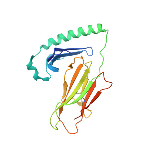
Crystal Structure of Hla-Dq0602 that Protects Against Type 1 Diabetes and Confers Strong Susceptibility to Narcolepsy

Siebold, C.Hansen, B.E.Wyer, J.R.Harlos, K.Esnouf, R.E.Svejgaard, A.Bell, J.I.Strominger, J.L.Jones, E.Y.Fugger, L.

(2004) Proc Natl Acad Sci U S A 101: 1999

  • DOI: https://doi.org/10.1073/pnas.0308458100
  • Primary Citation of Related Structures:  
    1UVQ

  • PubMed Abstract: 
  • The MHC class II molecule DQ0602 confers strong susceptibility to narcolepsy but dominant protection against type 1 diabetes. The crystal structure of DQ0602 reveals the molecular features underlying these contrasting genetic properties. Structural comparisons to homologous DQ molecules with differential disease associations highlight a previously unrecognized interplay between the volume of the P6 pocket and the specificity of the P9 pocket, which implies that presentation of an expanded peptide repertoire is critical for dominant protection against type 1 diabetes ...

    The MHC class II molecule DQ0602 confers strong susceptibility to narcolepsy but dominant protection against type 1 diabetes. The crystal structure of DQ0602 reveals the molecular features underlying these contrasting genetic properties. Structural comparisons to homologous DQ molecules with differential disease associations highlight a previously unrecognized interplay between the volume of the P6 pocket and the specificity of the P9 pocket, which implies that presentation of an expanded peptide repertoire is critical for dominant protection against type 1 diabetes. In narcolepsy, the volume of the P4 pocket appears central to the susceptibility, suggesting that the presentation of a specific peptide population plays a major role.


    Organizational Affiliation

    Division of Structural Biology, The Henry Wellcome Building for Genomic Medicine, University of Oxford, Roosevelt Drive, Oxford OX3 7BN, United Kingdom.


Macromolecules
Find similar proteins by:  (by identity cutoff)  |  3D Structure
Entity ID: 1
MoleculeChainsSequence LengthOrganismDetailsImage
HLA CLASS II HISTOCOMPATIBILITY ANTIGEN197Homo sapiensMutation(s): 0 
Gene Names: HLA-DQA1
UniProt
Find proteins for E9PMV2 (Homo sapiens)
Explore E9PMV2 
Go to UniProtKB:  E9PMV2
Entity Groups  
Sequence Clusters30% Identity50% Identity70% Identity90% Identity95% Identity100% Identity
UniProt GroupE9PMV2
Protein Feature View
Expand
  • Reference Sequence
Find similar proteins by:  (by identity cutoff)  |  3D Structure
Entity ID: 2
MoleculeChainsSequence LengthOrganismDetailsImage
HLA CLASS II HISTOCOMPATIBILITY ANTIGEN198Homo sapiensMutation(s): 0 
Gene Names: HLA-DQB1HLA-DQB
UniProt & NIH Common Fund Data Resources
Find proteins for P01920 (Homo sapiens)
Explore P01920 
Go to UniProtKB:  P01920
PHAROS:  P01920
Entity Groups  
Sequence Clusters30% Identity50% Identity70% Identity90% Identity95% Identity100% Identity
UniProt GroupP01920
Protein Feature View
Expand
  • Reference Sequence
Find similar proteins by:  (by identity cutoff)  |  3D Structure
Entity ID: 3
MoleculeChainsSequence LengthOrganismDetailsImage
OREXIN33Homo sapiensMutation(s): 0 
Gene Names: HCRTOXPPORXPPOX
UniProt & NIH Common Fund Data Resources
Find proteins for O43612 (Homo sapiens)
Explore O43612 
Go to UniProtKB:  O43612
PHAROS:  O43612
Entity Groups  
Sequence Clusters30% Identity50% Identity70% Identity90% Identity95% Identity100% Identity
UniProt GroupO43612
Protein Feature View
Expand
  • Reference Sequence
Oligosaccharides

Help

Entity ID: 4
MoleculeChainsChain Length2D DiagramGlycosylation3D Interactions
beta-D-mannopyranose-(1-4)-2-acetamido-2-deoxy-beta-D-glucopyranose-(1-4)-[alpha-L-fucopyranose-(1-6)]2-acetamido-2-deoxy-beta-D-glucopyranose
D
4N-Glycosylation Oligosaccharides Interaction
Glycosylation Resources
GlyTouCan:  G32152BH
GlyCosmos:  G32152BH
GlyGen:  G32152BH
Small Molecules
Ligands 4 Unique
IDChainsName / Formula / InChI Key2D Diagram3D Interactions
NAG
Query on NAG

Download Ideal Coordinates CCD File 
F [auth A],
G [auth A]
2-acetamido-2-deoxy-beta-D-glucopyranose
C8 H15 N O6
OVRNDRQMDRJTHS-FMDGEEDCSA-N
 Ligand Interaction
GLY
Query on GLY

Download Ideal Coordinates CCD File 
E [auth A]GLYCINE
C2 H5 N O2
DHMQDGOQFOQNFH-UHFFFAOYSA-N
 Ligand Interaction
ZN
Query on ZN

Download Ideal Coordinates CCD File 
H [auth B]ZINC ION
Zn
PTFCDOFLOPIGGS-UHFFFAOYSA-N
 Ligand Interaction
ACY
Query on ACY

Download Ideal Coordinates CCD File 
I [auth B]ACETIC ACID
C2 H4 O2
QTBSBXVTEAMEQO-UHFFFAOYSA-N
 Ligand Interaction
Experimental Data & Validation

Experimental Data

  • Method: X-RAY DIFFRACTION
  • Resolution: 1.80 Å
  • R-Value Free: 0.205 
  • R-Value Work: 0.189 
  • R-Value Observed: 0.189 
  • Space Group: P 21 21 2
Unit Cell:
Length ( Å )Angle ( ˚ )
a = 102.078α = 90
b = 129.304β = 90
c = 40.619γ = 90
Software Package:
Software NamePurpose
DENZOdata reduction
SCALEPACKdata scaling
EPMRphasing
CNSphasing
CNSrefinement

Structure Validation

View Full Validation Report


Ligand Structure Quality Assessment 


Entry History 

Deposition Data

Revision History  (Full details and data files)

  • Version 1.0: 2004-02-05
    Type: Initial release
  • Version 1.1: 2015-10-21
    Changes: Derived calculations, Non-polymer description, Other, Refinement description, Source and taxonomy, Structure summary, Version format compliance
  • Version 2.0: 2020-07-29
    Type: Remediation
    Reason: Carbohydrate remediation
    Changes: Advisory, Atomic model, Data collection, Derived calculations, Other, Structure summary