Elucidating the specificity of binding of sulfonylurea herbicides to acetohydroxyacid synthase.
McCourt, J.A., Pang, S.S., Guddat, L.W., Duggleby, R.G.(2005) Biochemistry 44: 2330-2338
- PubMed: 15709745 Search on PubMed
- DOI: https://doi.org/10.1021/bi047980a
- Primary Citation Related Structures:
1T9A, 1T9B, 1T9C, 1T9D - PubMed Abstract:
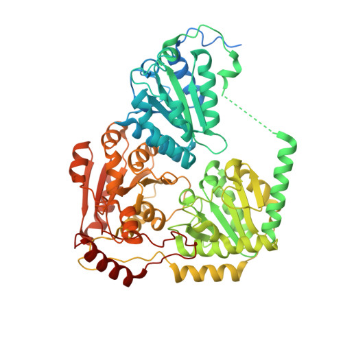
Acetohydroxyacid synthase (AHAS, EC 2.2.1.6) is the target for the sulfonylurea herbicides, which act as potent inhibitors of the enzyme. Chlorsulfuron (marketed as Glean) and sulfometuron methyl (marketed as Oust) are two commercially important members of this family of herbicides. Here we report crystal structures of yeast AHAS in complex with chlorsulfuron (at a resolution of 2.19 A), sulfometuron methyl (2.34 A), and two other sulfonylureas, metsulfuron methyl (2.29 A) and tribenuron methyl (2.58 A). The structures observed suggest why these inhibitors have different potencies and provide clues about the differential effects of mutations in the active site tunnel on various inhibitors. In all of the structures, the thiamin diphosphate cofactor is fragmented, possibly as the result of inhibitor binding. In addition to thiamin diphosphate, AHAS requires FAD for activity. Recently, it has been reported that reduction of FAD can occur as a minor side reaction due to reaction with the carbanion/enamine of the hydroxyethyl-ThDP intermediate that is formed midway through the catalytic cycle. Here we report that the isoalloxazine ring has a bent conformation that would account for its ability to accept electrons from the hydroxyethyl intermediate. Most sequence and mutation data suggest that yeast AHAS is a high-quality model for the plant enzyme.
- Department of Biochemistry and Molecular Biology, The University of Queensland, Brisbane, Queensland 4072, Australia.
Organizational Affiliation: