1T7W

Zn-alpha-2-glycoprotein; CHO-ZAG PEG 400


Experimental Data Snapshot

  • Method: X-RAY DIFFRACTION
  • Resolution: 2.70 Å
  • R-Value Free: 0.290 
  • R-Value Work: 0.232 

Starting Model: experimental
View more details

wwPDB Validation   3D Report Full Report


Ligand Structure Quality Assessment 


This is version 2.2 of the entry. See complete history


Literature

Crystallographic studies of ligand binding by Zn-alpha2-glycoprotein.

Delker, S.L.West Jr., A.P.McDermott, L.Kennedy, M.W.Bjorkman, P.J.

(2004) J Struct Biol 148: 205-213

  • DOI: https://doi.org/10.1016/j.jsb.2004.04.009
  • Primary Citation of Related Structures:  
    1T7V, 1T7W, 1T7X, 1T7Y, 1T7Z, 1T80

  • PubMed Abstract: 

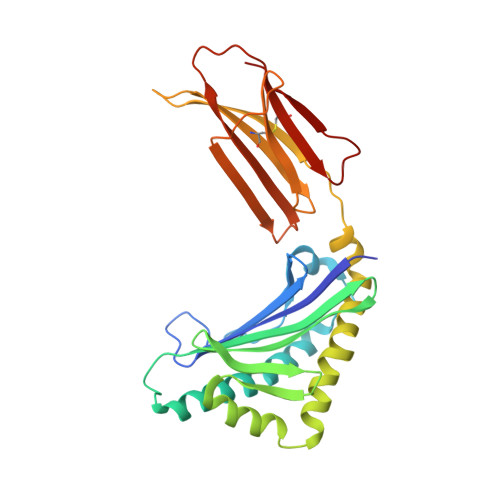
    Zn-alpha2-glycoprotein (ZAG) is a 41 kDa soluble protein that is present in most bodily fluids. The previously reported 2.8 A crystal structure of ZAG isolated from human serum demonstrated the structural similarity between ZAG and class I major histocompatibility complex (MHC) molecules and revealed a non-peptidic ligand in the ZAG counterpart of the MHC peptide-binding groove. Here we present crystallographic studies to explore further the nature of the non-peptidic ligand in the ZAG groove. Comparison of the structures of several forms of recombinant ZAG, including a 1.95 A structure derived from ZAG expressed in insect cells, suggests that the non-peptidic ligand in the current structures and in the structure of serum ZAG is a polyethylene glycol (PEG), which is present in the crystallization conditions used. Further support for PEG binding in the ZAG groove is provided by the finding that PEG displaces a fluorophore-tagged fatty acid from the ZAG binding site. From these results we hypothesize that our purified forms of ZAG do not contain a bound endogenous ligand, but that the ZAG groove is capable of binding hydrophobic molecules, which may relate to its function.


  • Organizational Affiliation

    Division of Biology, 114-96 Pasadena, CA 91125, USA.


Macromolecules
Find similar proteins by:  (by identity cutoff)  |  3D Structure
Entity ID: 1
MoleculeChains Sequence LengthOrganismDetailsImage
Zinc-alpha-2-glycoprotein278Homo sapiensMutation(s): 2 
Gene Names: AZGP1ZAGZNGP1
UniProt & NIH Common Fund Data Resources
Find proteins for P25311 (Homo sapiens)
Explore P25311 
Go to UniProtKB:  P25311
GTEx:  ENSG00000160862 
Entity Groups  
Sequence Clusters30% Identity50% Identity70% Identity90% Identity95% Identity100% Identity
UniProt GroupP25311
Glycosylation
Glycosylation Sites: 2Go to GlyGen: P25311-1
Sequence Annotations
Expand
  • Reference Sequence
Oligosaccharides

Help

Entity ID: 2
MoleculeChains Length2D Diagram Glycosylation3D Interactions
2-acetamido-2-deoxy-beta-D-glucopyranose-(1-4)-2-acetamido-2-deoxy-beta-D-glucopyranose
B
2N-Glycosylation
Glycosylation Resources
GlyTouCan:  G42666HT
GlyCosmos:  G42666HT
GlyGen:  G42666HT
Small Molecules
Ligands 1 Unique
IDChains Name / Formula / InChI Key2D Diagram3D Interactions
NAG
Query on NAG

Download Ideal Coordinates CCD File 
C [auth A]2-acetamido-2-deoxy-beta-D-glucopyranose
C8 H15 N O6
OVRNDRQMDRJTHS-FMDGEEDCSA-N
Experimental Data & Validation

Experimental Data

  • Method: X-RAY DIFFRACTION
  • Resolution: 2.70 Å
  • R-Value Free: 0.290 
  • R-Value Work: 0.232 
  • Space Group: P 43 21 2
Unit Cell:
Length ( Å )Angle ( ˚ )
a = 121.681α = 90
b = 121.681β = 90
c = 65.36γ = 90
Software Package:
Software NamePurpose
CNSrefinement
DENZOdata reduction
SCALEPACKdata scaling
AMoREphasing

Structure Validation

View Full Validation Report


Ligand Structure Quality Assessment 


Entry History 

Deposition Data

Revision History  (Full details and data files)

  • Version 1.0: 2004-12-21
    Type: Initial release
  • Version 1.1: 2008-04-30
    Changes: Version format compliance
  • Version 1.2: 2011-07-13
    Changes: Non-polymer description, Version format compliance
  • Version 1.3: 2017-10-11
    Changes: Refinement description
  • Version 2.0: 2020-07-29
    Type: Remediation
    Reason: Carbohydrate remediation
    Changes: Atomic model, Data collection, Derived calculations, Structure summary
  • Version 2.1: 2021-10-27
    Changes: Database references, Structure summary
  • Version 2.2: 2023-08-23
    Changes: Data collection, Refinement description