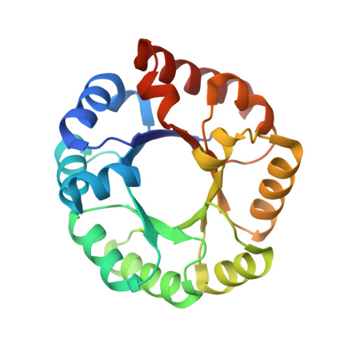
Evolution of Enzymatic Activities in the Orotidine 5'-Monophosphate Decarboxylase Suprafamily: Crystallographic Evidence for a Proton Relay System in the Active Site of 3-Keto-l-gulonate 6-Phosphate Decarboxylase(,)
Wise, E.L., Yew, W.S., Gerlt, J.A., Rayment, I.(2004) Biochemistry 43: 6438-6446
- PubMed: 15157078 Search on PubMed
- DOI: https://doi.org/10.1021/bi0497392
- Primary Citation Related Structures:
1SO3, 1SO4, 1SO5, 1SO6 - PubMed Abstract:
3-Keto-L-gulonate 6-phosphate decarboxylase (KGPDC), a member of the orotidine monophosphate decarboxylase (OMPDC) suprafamily, catalyzes the Mg(2+)-dependent decarboxylation of 3-keto-L-gulonate 6-phosphate to L-xylulose 5-phosphate. Structural and biochemical evidence suggests that the KGPDC reaction proceeds via a Mg(2+)-stabilized 1,2-cis-enediolate intermediate. Protonation of the enediolate intermediate occurs in a nonstereospecific manner to form L-xylulose 5-phosphate. Although the exact mechanism of proton delivery is not known, Glu112, His136, and Arg139 have been implicated in this process [Yew, W. S., Wise, E., Rayment, I., and Gerlt, J. A. (2004) Biochemistry 43, 6427-6437]. Surprisingly, single amino acid substitutions of these positions do not substantially reduce catalytic activity but rather alter the stereochemical course of the reaction. Here, we report the X-ray crystal structures of four mutants, K64A, H136A, E112Q, and E112Q/H136A, each determined in the presence of L-threonohydroxamate 4-phosphate, an analogue of the enediolate intermediate, to 1.7, 1.9, 1.8, and 1.9 A resolution, respectively. These structures reveal that substitutions of Lys64, Glu112, and His136 cause changes in the positions of the intermediate analogue and two active site water molecules that were previously identified as possible proton donors. These changes correlate with the observed alterations in the reaction stereochemistry for these mutants, thereby supporting a reaction mechanism in which water molecules competitively shuttle protons from the side chains of His136 and Arg139 to alternate faces of the cis-enediolate intermediate. These studies further underscore the wide variation in the reaction mechanisms in the OMPDC suprafamily.
- Department of Biochemistry, University of Wisconsin, Madison, Wisconsin 53706, USA.
Organizational Affiliation: