1P0I

Crystal structure of human butyryl cholinesterase


Experimental Data Snapshot

  • Method: X-RAY DIFFRACTION
  • Resolution: 2.00 Å
  • R-Value Free: 0.225 
  • R-Value Work: 0.195 
  • R-Value Observed: 0.195 

wwPDB Validation   3D Report Full Report


Ligand Structure Quality Assessment 


This is version 2.1 of the entry. See complete history


Literature

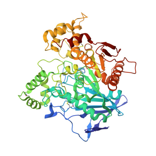
Crystal structure of human butyrylcholinesterase and of its complexes with substrate and products.

Nicolet, Y.Lockridge, O.Masson, P.Fontecilla-Camps, J.C.Nachon, F.

(2003) J Biol Chem 278: 41141-41147

  • DOI: 10.1074/jbc.M210241200
  • Primary Citation of Related Structures:  
    1P0I, 1P0M, 1P0P, 1P0Q

  • PubMed Abstract: 
  • Cholinesterases are among the most efficient enzymes known. They are divided into two groups: acetylcholinesterase, involved in the hydrolysis of the neurotransmitter acetylcholine, and butyrylcholinesterase of unknown function. Several crystal structures of the former have shown that the active site is located at the bottom of a deep and narrow gorge, raising the question of how substrate and products enter and leave ...

    Cholinesterases are among the most efficient enzymes known. They are divided into two groups: acetylcholinesterase, involved in the hydrolysis of the neurotransmitter acetylcholine, and butyrylcholinesterase of unknown function. Several crystal structures of the former have shown that the active site is located at the bottom of a deep and narrow gorge, raising the question of how substrate and products enter and leave. Human butyrylcholinesterase (BChE) has attracted attention because it can hydrolyze toxic esters such as cocaine or scavenge organophosphorus pesticides and nerve agents. Here we report the crystal structures of several recombinant truncated human BChE complexes and conjugates and provide a description for mechanistically relevant non-productive substrate and product binding. As expected, the structure of BChE is similar to a previously published theoretical model of this enzyme and to the structure of Torpedo acetylcholinesterase. The main difference between the experimentally determined BChE structure and its model is found at the acyl binding pocket that is significantly bigger than expected. An electron density peak close to the catalytic Ser(198) has been modeled as bound butyrate.


    Related Citations: 
    • Engineering of a monomeric and low-glycosylated form of human butyrylcholinesterase: expression, purification, characterization and crystallization
      Nachon, F., Nicolet, Y., Viguie, N., Masson, P., Fontecilla-Camps, J.C., Lockridge, O.
      (2002) Eur J Biochem 269: 630

    Organizational Affiliation

    Laboratoire de Cristallographie et Cristallogénèse des Protéines, Institut de Biologie Structurale "Jean-Pierre Ebel," CEA, UJF, CNRS, 41 rue Jules Horowitz, 38027 Grenoble Cedex 1, France.


Macromolecules
Find similar proteins by:  (by identity cutoff)  |  3D Structure
Entity ID: 1
MoleculeChainsSequence LengthOrganismDetailsImage
CholinesteraseA529Homo sapiensMutation(s): 4 
Gene Names: BCHE OR CHE1BCHECHE1
EC: 3.1.1.8
Membrane Entity: Yes 
UniProt & NIH Common Fund Data Resources
Find proteins for P06276 (Homo sapiens)
Explore P06276 
Go to UniProtKB:  P06276
PHAROS:  P06276
Entity Groups  
Sequence Clusters30% Identity50% Identity70% Identity90% Identity95% Identity100% Identity
UniProt GroupP06276
Protein Feature View
Expand
  • Reference Sequence
Oligosaccharides

Help

Entity ID: 2
MoleculeChainsChain Length2D DiagramGlycosylation3D Interactions
2-acetamido-2-deoxy-beta-D-glucopyranose-(1-4)-[alpha-L-fucopyranose-(1-6)]2-acetamido-2-deoxy-beta-D-glucopyranoseB, C 3N-Glycosylation Oligosaccharides Interaction
Glycosylation Resources
GlyTouCan:  G21290RB
GlyCosmos:  G21290RB
GlyGen:  G21290RB
Small Molecules
Ligands 6 Unique
IDChainsName / Formula / InChI Key2D Diagram3D Interactions
NAG
Query on NAG

Download Ideal Coordinates CCD File 
D [auth A],
E [auth A],
F [auth A]
2-acetamido-2-deoxy-beta-D-glucopyranose
C8 H15 N O6
OVRNDRQMDRJTHS-FMDGEEDCSA-N
 Ligand Interaction
MES
Query on MES

Download Ideal Coordinates CCD File 
L [auth A]2-(N-MORPHOLINO)-ETHANESULFONIC ACID
C6 H13 N O4 S
SXGZJKUKBWWHRA-UHFFFAOYSA-N
 Ligand Interaction
SO4
Query on SO4

Download Ideal Coordinates CCD File 
G [auth A],
H [auth A]
SULFATE ION
O4 S
QAOWNCQODCNURD-UHFFFAOYSA-L
 Ligand Interaction
GOL
Query on GOL

Download Ideal Coordinates CCD File 
M [auth A],
N [auth A],
O [auth A]
GLYCEROL
C3 H8 O3
PEDCQBHIVMGVHV-UHFFFAOYSA-N
 Ligand Interaction
BUA
Query on BUA

Download Ideal Coordinates CCD File 
K [auth A]butanoic acid
C4 H8 O2
FERIUCNNQQJTOY-UHFFFAOYSA-N
 Ligand Interaction
CL
Query on CL

Download Ideal Coordinates CCD File 
I [auth A],
J [auth A]
CHLORIDE ION
Cl
VEXZGXHMUGYJMC-UHFFFAOYSA-M
 Ligand Interaction
Experimental Data & Validation

Experimental Data

  • Method: X-RAY DIFFRACTION
  • Resolution: 2.00 Å
  • R-Value Free: 0.225 
  • R-Value Work: 0.195 
  • R-Value Observed: 0.195 
  • Space Group: I 4 2 2
Unit Cell:
Length ( Å )Angle ( ˚ )
a = 154.66α = 90
b = 154.66β = 90
c = 127.89γ = 90
Software Package:
Software NamePurpose
CNSrefinement
MOSFLMdata reduction
CCP4data scaling
AMoREphasing

Structure Validation

View Full Validation Report


Ligand Structure Quality Assessment 


Entry History 

Deposition Data

Revision History  (Full details and data files)

  • Version 1.0: 2003-08-05
    Type: Initial release
  • Version 1.1: 2008-04-29
    Changes: Version format compliance
  • Version 1.2: 2011-07-13
    Changes: Derived calculations, Version format compliance
  • Version 2.0: 2020-07-29
    Type: Remediation
    Reason: Carbohydrate remediation
    Changes: Advisory, Atomic model, Data collection, Derived calculations, Structure summary
  • Version 2.1: 2021-10-27
    Changes: Advisory, Database references, Derived calculations, Structure summary