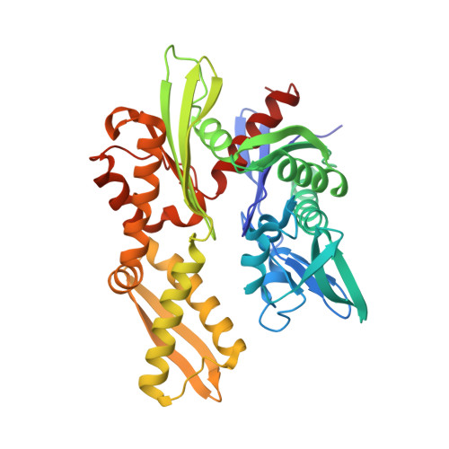
1NGJ

STRUCTURAL BASIS OF THE 70-KILODALTON HEAT SHOCK COGNATE PROTEIN ATP HYDROLYTIC ACTIVITY, II. STRUCTURE OF THE ACTIVE SITE WITH ADP OR ATP BOUND TO WILD TYPE AND MUTANT ATPASE FRAGMENT


Experimental Data Snapshot

  • Method: X-RAY DIFFRACTION
  • Resolution: 2.10 Å
  • R-Value Work: 0.194 
  • R-Value Observed: 0.194 

wwPDB Validation   3D Report Full Report


This is version 1.3 of the entry. See complete history


Literature

Structural basis of the 70-kilodalton heat shock cognate protein ATP hydrolytic activity. II. Structure of the active site with ADP or ATP bound to wild type and mutant ATPase fragment.

Flaherty, K.M.Wilbanks, S.M.DeLuca-Flaherty, C.McKay, D.B.

(1994) J Biol Chem 269: 12899-12907

  • Primary Citation of Related Structures:  
    1NGA, 1NGB, 1NGC, 1NGD, 1NGE, 1NGF, 1NGG, 1NGH, 1NGI, 1NGJ

  • PubMed Abstract: 

    The ATPase fragment of the bovine 70-kDa heat shock cognate protein is an attractive construct in which to study its mechanism of ATP hydrolysis. The three-dimensional structure suggests several residues that might participate in the ATPase reaction. Four acidic residues (Asp-10, Glu-175, Asp-199, and Asp-206) have been individually mutated to both the cognate amine (asparagine/glutamine) and to serine, and the effects of the mutations on the kinetics of the ATPase activity (Wilbanks, S. M., DeLuca-Flaherty, C., and McKay, D. B. (1994) J. Biol. Chem. 269, 12893-12898) and the structure of the mutant ATPase fragments have been determined, typically to approximately 2.4 A resolution. Additionally, the structures of the wild type protein complexed with MgADP and Pi, MgAMPPNP (5'-adenylyl-beta, gamma-imidodiphosphate) and CaAMPPNP have been refined to 2.1, 2.4, and 2.4 A, respectively. Combined, these structures provide models for the prehydrolysis, MgATP-bound state and the post-hydrolysis, MgADP-bound state of the ATPase fragment. These models suggest a pathway for the hydrolytic reaction in which 1) the gamma phosphate of bound ATP reorients to form a beta, gamma-bidentate phosphate complex with the Mg2+ ion, allowing 2) in-line nucleophilic attack on the gamma phosphate by a H2O molecule or OH- ion, with 3) subsequent release of inorganic phosphate.


  • Organizational Affiliation

    Beckman Laboratories for Structural Biology, Department of Cell Biology, Stanford University School of Medicine, California 94305.


Macromolecules
Find similar proteins by:  (by identity cutoff)  |  3D Structure
Entity ID: 1
MoleculeChains Sequence LengthOrganismDetailsImage
HEAT-SHOCK COGNATE 70 kD PROTEIN386Bos taurusMutation(s): 0 
EC: 3.6.1.3
UniProt
Find proteins for P19120 (Bos taurus)
Explore P19120 
Go to UniProtKB:  P19120
Entity Groups  
Sequence Clusters30% Identity50% Identity70% Identity90% Identity95% Identity100% Identity
UniProt GroupP19120
Sequence Annotations
Expand
  • Reference Sequence
Small Molecules
Ligands 2 Unique
IDChains Name / Formula / InChI Key2D Diagram3D Interactions
ANP
Query on ANP

Download Ideal Coordinates CCD File 
C [auth A]PHOSPHOAMINOPHOSPHONIC ACID-ADENYLATE ESTER
C10 H17 N6 O12 P3
PVKSNHVPLWYQGJ-KQYNXXCUSA-N
MG
Query on MG

Download Ideal Coordinates CCD File 
B [auth A]MAGNESIUM ION
Mg
JLVVSXFLKOJNIY-UHFFFAOYSA-N
Experimental Data & Validation

Experimental Data

  • Method: X-RAY DIFFRACTION
  • Resolution: 2.10 Å
  • R-Value Work: 0.194 
  • R-Value Observed: 0.194 
  • Space Group: P 21 21 21
Unit Cell:
Length ( Å )Angle ( ˚ )
a = 145.3α = 90
b = 65β = 90
c = 46.9γ = 90
Software Package:
Software NamePurpose
X-PLORmodel building
X-PLORrefinement
X-PLORphasing

Structure Validation

View Full Validation Report


Entry History 

Deposition Data

Revision History  (Full details and data files)

  • Version 1.0: 1994-08-31
    Type: Initial release
  • Version 1.1: 2008-03-24
    Changes: Version format compliance
  • Version 1.2: 2011-07-13
    Changes: Version format compliance
  • Version 1.3: 2024-02-14
    Changes: Data collection, Database references, Derived calculations, Other