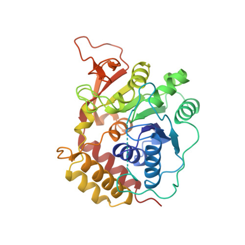
1MAS

PURINE NUCLEOSIDE HYDROLASE


Experimental Data Snapshot

  • Method: X-RAY DIFFRACTION
  • Resolution: 2.50 Å
  • R-Value Work: 0.170 

wwPDB Validation   3D Report Full Report


This is version 1.2 of the entry. See complete history


Literature

Three-dimensional structure of the inosine-uridine nucleoside N-ribohydrolase from Crithidia fasciculata.

Degano, M.Gopaul, D.N.Scapin, G.Schramm, V.L.Sacchettini, J.C.

(1996) Biochemistry 35: 5971-5981

  • DOI: 10.1021/bi952999m
  • Primary Citation of Related Structures:  
    1MAS

  • PubMed Abstract: 
  • Protozoan parasites rely on the host for purines since they lack a de novo synthetic pathway. Crithidia fasciculata salvages exogenous inosine primarily through hydrolysis of the N-ribosidic bond using several nucleoside hydrolases. The most abundant nucleoside hydrolase is relatively nonspecific but prefers inosine and uridine as substrates ...

    Protozoan parasites rely on the host for purines since they lack a de novo synthetic pathway. Crithidia fasciculata salvages exogenous inosine primarily through hydrolysis of the N-ribosidic bond using several nucleoside hydrolases. The most abundant nucleoside hydrolase is relatively nonspecific but prefers inosine and uridine as substrates. Here we report the three-dimensional structure of the inosine-uridine nucleoside hydrolase (IU-NH) from C. fasciculata determined by X-ray crystallography at a nominal resolution of 2.5 A. The enzyme has an open (alpha, beta) structure which differs from the classical dinucleotide binding fold. IU-nucleoside hydrolase is composed of a mixed eight-stranded beta sheet surrounded by six alpha helices and a small C-terminal lobe composed of four alpha helices. Two short antiparallel beta strands are involved in intermolecular contacts. The catalytic pocket is located at the C-terminal end of beta strands beta 1 and beta 4. Four aspartate residues are located at the bottom of the cavity in a geometry which suggests interaction with the ribose moiety of the nucleoside. These groups could provide the catalytically important interactions to the ribosyl hydroxyls and the stabilizing anion for the oxycarbonium-like transition state. Histidine 241, located on the side of the active site cavity, is the proposed proton donor which facilitates purine base departure [Gopaul, D. N., Meyer, S. L., Degano, M., Sacchettini, J. C., & Schramm, V. L. (1996) Biochemistry 35, 5963-5970]. The substrate binding site is unlike that from purine nucleoside phosphorylase, phosphoribosyltransferases, or uracil DNA glycosylase and thus represents a novel architecture for general acid-base catalysis. This detailed knowledge of the architecture of the active site, together with the previous transition state analysis [Horenstein, B. A., Parkin, D. W., Estupiñán, B., & Schramm, V. L. (1991) Biochemistry 30, 10788-10795], allows analysis of the interactions leading to catalysis and an explanation for the tight-binding inhibitors of the enzyme [Schramm, V. L., Horenstein, B. A., & Kline, P. C. (1994) J. Biol. Chem. 269, 18259-18262].


    Related Citations: 
    • Inosine-Uridine Nucleoside Hydrolase from Crithidia Fasciculata. Genetic Characterization, Crystallization, and Identification of Histidine 241 as a Catalytic Site Residue
      Gopaul, D.N., Meyer, S.L., Degano, M., Sacchettini, J.C., Schramm, V.L.
      (1996) Biochemistry 35: 5963
    • Transition-State Analysis of Nucleoside Hydrolase from Crithidia Fasciculata
      Horenstein, B.A., Parkin, D.W., Estupinan, B., Schramm, V.L.
      (1991) Biochemistry 30: 10788
    • Nucleoside Hydrolase from Crithidia Fasciculata
      Parkin, D.W., Horenstein, B.A., Abdulah, D.R., Estupinan, B., Schramm, V.L.
      (1991) J Biol Chem 266: 20658

    Organizational Affiliation

    Department of Biochemistry, Albert Einstein College of Medicine, Bronx, New York 10461, USA.


Macromolecules
Find similar proteins by:  (by identity cutoff)  |  3D Structure
Entity ID: 1
MoleculeChainsSequence LengthOrganismDetailsImage
INOSINE-URIDINE NUCLEOSIDE N-RIBOHYDROLASEA, B314Crithidia fasciculataMutation(s): 0 
Gene Names: IU-NH FROM C.FASCICULATAIUNH
EC: 3.2.2.1 (PDB Primary Data), 3.2.2.2 (UniProt), 3.2.2.3 (UniProt)
UniProt
Find proteins for Q27546 (Crithidia fasciculata)
Explore Q27546 
Go to UniProtKB:  Q27546
Entity Groups  
Sequence Clusters30% Identity50% Identity70% Identity90% Identity95% Identity100% Identity
UniProt GroupQ27546
Protein Feature View
Expand
  • Reference Sequence
Small Molecules
Ligands 1 Unique
IDChainsName / Formula / InChI Key2D Diagram3D Interactions
K
Query on K

Download Ideal Coordinates CCD File 
C [auth A],
D [auth B]
POTASSIUM ION
K
NPYPAHLBTDXSSS-UHFFFAOYSA-N
 Ligand Interaction
Experimental Data & Validation

Experimental Data

  • Method: X-RAY DIFFRACTION
  • Resolution: 2.50 Å
  • R-Value Work: 0.170 
  • Space Group: P 21 21 2
Unit Cell:
Length ( Å )Angle ( ˚ )
a = 63.84α = 90
b = 131.53β = 90
c = 90.1γ = 90
Software Package:
Software NamePurpose
X-PLORmodel building
TNTrefinement
X-PLORrefinement
XENGENdata reduction
X-PLORphasing

Structure Validation

View Full Validation Report


Entry History 

Deposition Data

Revision History  (Full details and data files)

  • Version 1.0: 1996-08-17
    Type: Initial release
  • Version 1.1: 2008-03-24
    Changes: Version format compliance
  • Version 1.2: 2011-07-13
    Changes: Version format compliance