1LO0 | pdb_00001lo0

Catalytic Retro-Diels-Alderase Transition State Analogue Complex


Experimental Data Snapshot

  • Method: X-RAY DIFFRACTION
  • Resolution: 2.00 Å
  • R-Value Free: 
    0.255 (Depositor) 
  • R-Value Work: 
    0.206 (Depositor) 
  • R-Value Observed: 
    0.206 (Depositor) 

Starting Model: experimental
View more details

wwPDB Validation 3D Report Full Report

Validation slider image for 1LO0

This is version 1.4 of the entry. See complete history

Literature

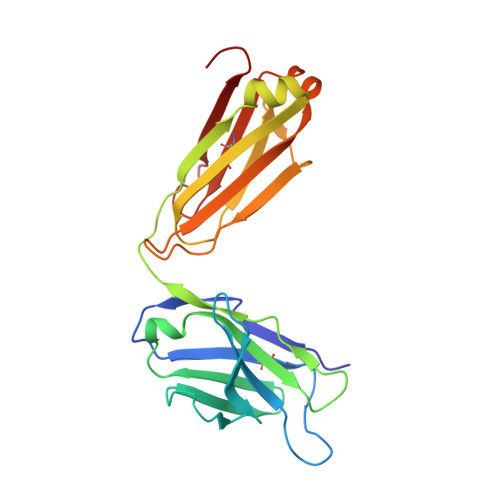
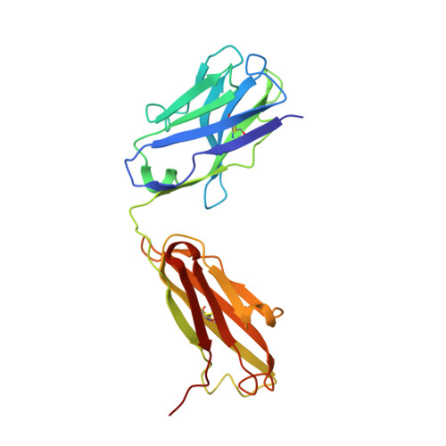
A structural basis for the activity of retro-Diels-Alder catalytic antibodies: evidence for a catalytic aromatic residue.

Hugot, M.Bensel, N.Vogel, M.Reymond, M.T.Stadler, B.Reymond, J.L.Baumann, U.

(2002) Proc Natl Acad Sci U S A 99: 9674-9678

  • DOI: https://doi.org/10.1073/pnas.142286599
  • Primary Citation Related Structures: 
    1LO0, 1LO2, 1LO3, 1LO4

  • PubMed Abstract: 

    The nitroxyl synthase catalytic antibodies 10F11, 9D9, and 27C5 catalyze the release of nitroxyl from a bicyclic pro-drug by accelerating a retro-Diels-Alder reaction. The Fabs (antigen-binding fragments) of these three catalytic antibodies were cloned and sequenced. Fab 9D9 was crystallized in the apo-form and in complex with one transition state analogue of the reaction. Crystal structures of Fab 10F11 in complex with ligands mimicking substrate, transition state, and product have been determined at resolutions ranging from 1.8 to 2.3 A. Antibodies 9D9 and 10F11 show increased shape complementarity (as quantified by the program sc) to the hapten and to a modeled transition state as compared with substrate and product. The shape complementarity is mediated to a large extent by an aromatic residue (tyrosine or tryptophan) at the bottom of the hydrophobic active pocket, which undergoes pi-stacking interactions with the aromatic rings of the ligands. Another factor contributing to the different reactivity of the regioisomers probably arises because of hydrogen-bonding interactions between the nitroxyl bridge and the backbone amide of PheH101 and possibly a conserved water molecule.


  • Organizational Affiliation
    • Department of Chemistry and Biochemistry, University of Berne, Freiestrasse 3, CH-3012 Berne, Switzerland.

Macromolecule Content 

  • Total Structure Weight: 96.62 kDa 
  • Atom Count: 6,905 
  • Modeled Residue Count: 878 
  • Deposited Residue Count: 878 
  • Unique protein chains: 2

Macromolecules

Find similar proteins by:|  3D Structure
Entity ID: 1
MoleculeChains  Sequence LengthOrganismDetailsImage
If kappa light chainA [auth X],
C [auth L]
219Mus musculusMutation(s): 0 
UniProt
Find proteins for P01837 (Mus musculus)
Explore P01837 
Go to UniProtKB:  P01837
Entity Groups
Sequence Clusters30% Identity50% Identity70% Identity90% Identity95% Identity100% Identity
UniProt GroupP01837
Sequence Annotations
Expand
Reference Sequence
Find similar proteins by:|  3D Structure
Entity ID: 2
MoleculeChains  Sequence LengthOrganismDetailsImage
Ig gamma 2a heavy chainB [auth Y],
D [auth H]
220Mus musculusMutation(s): 0 
UniProt
Find proteins for P01865 (Mus musculus)
Explore P01865 
Go to UniProtKB:  P01865
Entity Groups
Sequence Clusters30% Identity50% Identity70% Identity90% Identity95% Identity100% Identity
UniProt GroupP01865
Sequence Annotations
Expand
Reference Sequence

Small Molecules

Ligands 1 Unique
IDChains Name / Formula / InChI Key2D Diagram3D Interactions
BC1

Query on BC1


Download:Ideal Coordinates CCD File
E [auth Y],
F [auth H]
3-{[(9-CYANO-9,10-DIHYDRO-10-METHYLACRIDIN-9-YL)CARBONYL]AMINO}PROPANOIC ACID
C19 H17 N3 O3
FRBAOMHMZCGBOJ-UHFFFAOYSA-N

Experimental Data & Validation

Experimental Data

  • Method: X-RAY DIFFRACTION
  • Resolution: 2.00 Å
  • R-Value Free:  0.255 (Depositor) 
  • R-Value Work:  0.206 (Depositor) 
  • R-Value Observed: 0.206 (Depositor) 
Space Group: P 1 21 1
Unit Cell:
Length ( Å )Angle ( ˚ )
a = 40.528α = 90
b = 140.095β = 90
c = 85.476γ = 90
Software Package:
Software NamePurpose
MOSFLMdata reduction
SCALAdata scaling
CNSrefinement
CCP4data scaling
CNSphasing

Structure Validation

View Full Validation Report


Entry History 

Deposition Data

Revision History  (Full details and data files)

  • Version 1.0: 2002-06-26
    Type: Initial release
  • Version 1.1: 2008-04-28
    Changes: Version format compliance
  • Version 1.2: 2011-07-13
    Changes: Version format compliance
  • Version 1.3: 2024-04-03
    Changes: Data collection, Database references, Derived calculations, Refinement description
  • Version 1.4: 2024-10-09
    Changes: Structure summary