1KY1

METHIONINE CORE MUTANT OF T4 LYSOZYME


Experimental Data Snapshot

  • Method: X-RAY DIFFRACTION
  • Resolution: 2.05 Å
  • R-Value Work: 0.165 

wwPDB Validation   3D Report Full Report


Ligand Structure Quality Assessment 


This is version 1.3 of the entry. See complete history


Literature

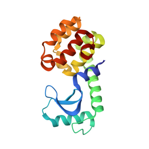
Multiple methionine substitutions are tolerated in T4 lysozyme and have coupled effects on folding and stability

Gassner, N.C.Baase, W.A.Mooers, B.H.Busam, R.D.Weaver, L.H.Lindstrom, J.D.Quillin, M.L.Matthews, B.W.

(2003) Biophys Chem 100: 325-340

  • DOI: 10.1016/s0301-4622(02)00290-9
  • Primary Citation of Related Structures:  
    1KS3, 1KW5, 1KW7, 1KY0, 1KY1, 1L0J, 1L0K, 1LPY, 1LW9, 1LWG, 1LWK

  • PubMed Abstract: 
  • In order to further explore the tolerance of proteins to amino acid substitutions within the interior, a series of core residues was replaced by methionine within the C-terminal domain of T4 lysozyme. By replacing leucine, isoleucine, valine and phenylalanine residues a total of 10 methionines could be introduced, which corresponds to a third of the residues that are buried in this domain ...

    In order to further explore the tolerance of proteins to amino acid substitutions within the interior, a series of core residues was replaced by methionine within the C-terminal domain of T4 lysozyme. By replacing leucine, isoleucine, valine and phenylalanine residues a total of 10 methionines could be introduced, which corresponds to a third of the residues that are buried in this domain. As more methionines are incorporated the protein gradually loses stability. This is attributed in part to a reduction in hydrophobic stabilization, in part to the increased entropic cost of localizing the long, flexible methionine sidechains, and in part to steric clashes. The changes in structure of the mutants relative to the wildtype protein are modest but tend to increase in an additive fashion as more methionines are included. In the most extreme case, namely the 10-methionine mutant, much of the C-terminal domain remains quite similar to wildtype (root-mean-square backbone shifts of 0.56 A), while the F and G helices undergo rotations of approximately 20 degrees and center-of-mass shifts of approximately 1.4 A. For up to six methionine substitutions the changes in stability are additive. Beyond this point, however, the multiple mutants are somewhat more stable than suggested from the sum of their constituents, especially for those including the replacement Val111-->Met. This is interpreted in terms of the larger structural changes associated with this substitution. The substituted sidechains in the mutant structures have somewhat higher crystallographic thermal factors than their counterparts in WT*. Nevertheless, the interiors of the mutant proteins retain a well-defined structure with little suggestion of molten-globule characteristics. Lysozymes in which selenomethionine has been incorporated rather than methionine tend to have increased stability. At the same time they also fold faster. This provides further evidence that, at the rate-limiting step in folding, the structure of the C-terminal domain of T4 lysozyme is similar to that of the fully folded protein.


    Related Citations: 
    • Methionine and alanine substitutions show that the formation of wild-type-like structure in the carboxy-terminal domain of T4 lysozymne is a rate-limiting step in folding
      Gassner, N.C., Baase, W.A., Lindstrom, J., Lu, J., Dahlquist, F.W., Matthews, B.W.
      (1999) Biochemistry 38: 14451
    • Structure of bacteriophage T4 lysozyme refined at 1.7 A resolution
      Weaver, L.H., Matthews, B.W.
      (1987) J Mol Biol 193: 189

    Organizational Affiliation

    Institute of Molecular Biology, Howard Hughes Medical Institute, 1229 University of Oregon, Eugene, OR 97403-1229, USA.


Macromolecules
Find similar proteins by:  (by identity cutoff)  |  3D Structure
Entity ID: 1
MoleculeChainsSequence LengthOrganismDetailsImage
LYSOZYMEA162Tequatrovirus T4Mutation(s): 8 
Gene Names: gene EE
EC: 3.2.1.17
UniProt
Find proteins for P00720 (Enterobacteria phage T4)
Explore P00720 
Go to UniProtKB:  P00720
Entity Groups  
Sequence Clusters30% Identity50% Identity70% Identity90% Identity95% Identity100% Identity
UniProt GroupP00720
Protein Feature View
Expand
  • Reference Sequence
Small Molecules
Ligands 2 Unique
IDChainsName / Formula / InChI Key2D Diagram3D Interactions
HED
Query on HED

Download Ideal Coordinates CCD File 
D [auth A]2-HYDROXYETHYL DISULFIDE
C4 H10 O2 S2
KYNFOMQIXZUKRK-UHFFFAOYSA-N
 Ligand Interaction
CL
Query on CL

Download Ideal Coordinates CCD File 
B [auth A],
C [auth A]
CHLORIDE ION
Cl
VEXZGXHMUGYJMC-UHFFFAOYSA-M
 Ligand Interaction
Experimental Data & Validation

Experimental Data

  • Method: X-RAY DIFFRACTION
  • Resolution: 2.05 Å
  • R-Value Work: 0.165 
  • Space Group: P 32 2 1
Unit Cell:
Length ( Å )Angle ( ˚ )
a = 61.4α = 90
b = 61.4β = 90
c = 96.5γ = 120
Software Package:
Software NamePurpose
STRATdata collection
AUTOSTRATdata reduction
TNTrefinement
STRATdata reduction
AUTOSTRATdata scaling
TNTphasing

Structure Validation

View Full Validation Report


Ligand Structure Quality Assessment 


Entry History 

Deposition Data

Revision History  (Full details and data files)

  • Version 1.0: 2003-06-03
    Type: Initial release
  • Version 1.1: 2008-04-28
    Changes: Version format compliance
  • Version 1.2: 2011-07-13
    Changes: Version format compliance
  • Version 1.3: 2021-10-27
    Changes: Database references, Derived calculations