Mechanistic basis for suicide inactivation of porphobilinogen synthase by 4,7-dioxosebacic acid, an inhibitor that shows dramatic species selectivity.
Kervinen, J., Jaffe, E.K., Stauffer, F., Neier, R., Wlodawer, A., Zdanov, A.(2001) Biochemistry 40: 8227-8236
- PubMed: 11444968 Search on PubMed
- DOI: https://doi.org/10.1021/bi010656k
- Primary Citation Related Structures:
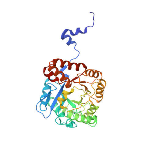
1I8J - PubMed Abstract:
4,7-Dioxosebacic acid (4,7-DOSA) is an active site-directed irreversible inhibitor of porphobilinogen synthase (PBGS). PBGS catalyzes the first common step in the biosynthesis of the tetrapyrrole cofactors such as heme, vitamin B(12), and chlorophyll. 4,7-DOSA was designed as an analogue of a proposed reaction intermediate in the physiological PBGS-catalyzed condensation of two molecules of 5-aminolevulinic acid. As shown here, 4,7-DOSA exhibits time-dependent and dramatic species-specific inhibition of PBGS enzymes. IC(50) values vary from 1 microM to 2.4 mM for human, Escherichia coli, Bradyrhizobium japonicum, Pseudomonas aeruginosa, and pea enzymes. Those PBGS utilizing a catalytic Zn(2+) are more sensitive to 4,7-DOSA than those that do not. Weak inhibition of a human mutant PBGS establishes that the inactivation by 4,7-DOSA requires formation of a Schiff base to a lysine that normally forms a Schiff base intermediate to one substrate molecule. A 1.9 A resolution crystal structure of E. coli PBGS complexed with 4,7-DOSA (PDB code ) shows one dimer per asymmetric unit and reveals that the inhibitor forms two Schiff base linkages with each monomer, one to the normal Schiff base-forming Lys-246 and the other to a universally conserved "perturbing" Lys-194 (E. coli numbering). This is the first structure to show inhibitor binding at the second of two substrate-binding sites.
- Institute for Cancer Research, Fox Chase Cancer Center, 7701 Burholme Avenue, Philadelphia, Pennsylvania 19111, USA.
Organizational Affiliation: