1F75

CRYSTAL STRUCTURE OF UNDECAPRENYL DIPHOSPHATE SYNTHASE FROM MICROCOCCUS LUTEUS B-P 26


Experimental Data Snapshot

  • Method: X-RAY DIFFRACTION
  • Resolution: 2.20 Å
  • R-Value Free: 0.246 
  • R-Value Work: 0.190 

wwPDB Validation   3D Report Full Report


This is version 1.4 of the entry. See complete history


Literature

Crystal structure of cis-prenyl chain elongating enzyme, undecaprenyl diphosphate synthase.

Fujihashi, M.Zhang, Y.W.Higuchi, Y.Li, X.Y.Koyama, T.Miki, K.

(2001) Proc Natl Acad Sci U S A 98: 4337-4342

  • DOI: https://doi.org/10.1073/pnas.071514398
  • Primary Citation of Related Structures:  
    1F75

  • PubMed Abstract: 

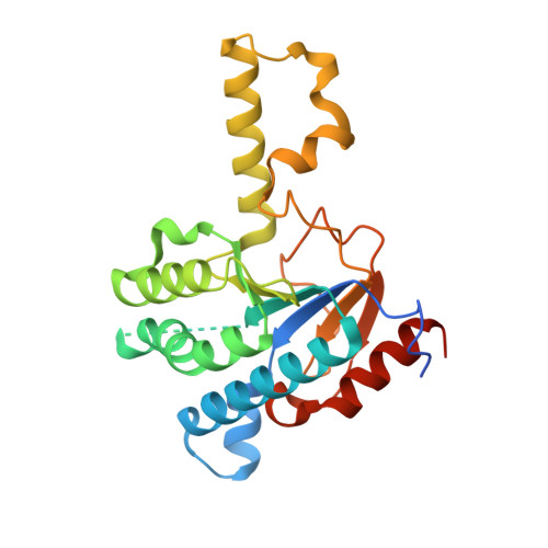
    Undecaprenyl diphosphate synthase (UPS) catalyzes the cis-prenyl chain elongation onto trans, trans-farnesyl diphosphate (FPP) to produce undecaprenyl diphosphate (UPP), which is indispensable for the biosynthesis of bacterial cell walls. We report here the crystal structure of UPS as the only three-dimensional structure among cis-prenyl chain elongating enzymes. The structure is classified into a protein fold family and is completely different from the so-called "isoprenoid synthase fold" that is believed to be a common structure for the enzymes relating to isoprenoid biosynthesis. Conserved amino acid residues among cis-prenyl chain elongating enzymes are located around a large hydrophobic cleft in the UPS structure. A structural P-loop motif, which frequently appears in the various kinds of phosphate binding site, is found at the entrance of this cleft. The catalytic site is determined on the basis of these structural features, from which a possible reaction mechanism is proposed.


  • Organizational Affiliation

    Department of Chemistry, Graduate School of Science, Kyoto University, Sakyo-ku, Kyoto 606-8502, Japan.


Macromolecules
Find similar proteins by:  (by identity cutoff)  |  3D Structure
Entity ID: 1
MoleculeChains Sequence LengthOrganismDetailsImage
UNDECAPRENYL PYROPHOSPHATE SYNTHETASE
A, B
249Micrococcus luteusMutation(s): 1 
EC: 2.5.1.31
UniProt
Find proteins for O82827 (Micrococcus luteus)
Explore O82827 
Go to UniProtKB:  O82827
Entity Groups  
Sequence Clusters30% Identity50% Identity70% Identity90% Identity95% Identity100% Identity
UniProt GroupO82827
Sequence Annotations
Expand
  • Reference Sequence
Small Molecules
Ligands 1 Unique
IDChains Name / Formula / InChI Key2D Diagram3D Interactions
SO4
Query on SO4

Download Ideal Coordinates CCD File 
C [auth A],
D [auth B]
SULFATE ION
O4 S
QAOWNCQODCNURD-UHFFFAOYSA-L
Experimental Data & Validation

Experimental Data

  • Method: X-RAY DIFFRACTION
  • Resolution: 2.20 Å
  • R-Value Free: 0.246 
  • R-Value Work: 0.190 
  • Space Group: C 1 2 1
Unit Cell:
Length ( Å )Angle ( ˚ )
a = 127.197α = 90
b = 60.161β = 105.69
c = 75.703γ = 90
Software Package:
Software NamePurpose
MLPHAREphasing
X-PLORrefinement
DENZOdata reduction
SCALEPACKdata scaling

Structure Validation

View Full Validation Report


Entry History 

Deposition Data

Revision History  (Full details and data files)

  • Version 1.0: 2001-03-28
    Type: Initial release
  • Version 1.1: 2008-04-27
    Changes: Version format compliance
  • Version 1.2: 2011-07-13
    Changes: Version format compliance
  • Version 1.3: 2021-11-03
    Changes: Database references, Derived calculations
  • Version 1.4: 2024-02-07
    Changes: Data collection