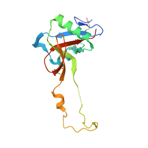
1EGG

STRUCTURE OF A C-TYPE CARBOHYDRATE-RECOGNITION DOMAIN (CRD-4) FROM THE MACROPHAGE MANNOSE RECEPTOR


Experimental Data Snapshot

  • Method: X-RAY DIFFRACTION
  • Resolution: 2.30 Å
  • R-Value Free: 0.278 
  • R-Value Work: 0.216 

wwPDB Validation   3D Report Full Report


This is version 1.2 of the entry. See complete history


Literature

Structure of a C-type carbohydrate recognition domain from the macrophage mannose receptor.

Feinberg, H.Park-Snyder, S.Kolatkar, A.R.Heise, C.T.Taylor, M.E.Weis, W.I.

(2000) J Biol Chem 275: 21539-21548

  • DOI: 10.1074/jbc.M002366200
  • Primary Citation of Related Structures:  
    1EGG, 1EGI

  • PubMed Abstract: 
  • The mannose receptor of macrophages and liver endothelium mediates clearance of pathogenic organisms and potentially harmful glycoconjugates. The extracellular portion of the receptor includes eight C-type carbohydrate recognition domains (CRDs), of which one, CRD-4, shows detectable binding to monosaccharide ligands ...

    The mannose receptor of macrophages and liver endothelium mediates clearance of pathogenic organisms and potentially harmful glycoconjugates. The extracellular portion of the receptor includes eight C-type carbohydrate recognition domains (CRDs), of which one, CRD-4, shows detectable binding to monosaccharide ligands. We have determined the crystal structure of CRD-4. Although the basic C-type lectin fold is preserved, a loop extends away from the core of the domain to form a domain-swapped dimer in the crystal. Of the two Ca(2+) sites, only the principal site known to mediate carbohydrate binding in other C-type lectins is occupied. This site is altered in a way that makes sugar binding impossible in the mode observed in other C-type lectins. The structure is likely to represent an endosomal form of the domain formed when Ca(2+) is lost from the auxiliary calcium site. The structure suggests a mechanism for endosomal ligand release in which the auxiliary calcium site serves as a pH sensor. Acid pH-induced removal of this Ca(2+) results in conformational rearrangements of the receptor, rendering it unable to bind carbohydrate ligands.


    Organizational Affiliation

    Department of Structural Biology, Stanford University School of Medicine, Stanford, California 94305, USA.


Macromolecules
Find similar proteins by:  (by identity cutoff)  |  3D Structure
Entity ID: 1
MoleculeChainsSequence LengthOrganismDetailsImage
MACROPHAGE MANNOSE RECEPTORA, B147Homo sapiensMutation(s): 0 
Gene Names: MRC1CLEC13DCLEC13DLMRC1L1
UniProt & NIH Common Fund Data Resources
Find proteins for P22897 (Homo sapiens)
Explore P22897 
Go to UniProtKB:  P22897
PHAROS:  P22897
Entity Groups  
Sequence Clusters30% Identity50% Identity70% Identity90% Identity95% Identity100% Identity
UniProt GroupP22897
Protein Feature View
Expand
  • Reference Sequence
Small Molecules
Ligands 1 Unique
IDChainsName / Formula / InChI Key2D Diagram3D Interactions
CA
Query on CA

Download Ideal Coordinates CCD File 
C [auth A],
D [auth A],
E [auth B]
CALCIUM ION
Ca
BHPQYMZQTOCNFJ-UHFFFAOYSA-N
 Ligand Interaction
Experimental Data & Validation

Experimental Data

  • Method: X-RAY DIFFRACTION
  • Resolution: 2.30 Å
  • R-Value Free: 0.278 
  • R-Value Work: 0.216 
  • Space Group: C 1 2 1
Unit Cell:
Length ( Å )Angle ( ˚ )
a = 129.89α = 90
b = 38.28β = 99.09
c = 54.81γ = 90
Software Package:
Software NamePurpose
DENZOdata reduction
SCALEPACKdata scaling
AMoREphasing
CNSrefinement

Structure Validation

View Full Validation Report


Entry History 

Deposition Data

Revision History  (Full details and data files)

  • Version 1.0: 2000-08-30
    Type: Initial release
  • Version 1.1: 2008-04-27
    Changes: Version format compliance
  • Version 1.2: 2011-07-13
    Changes: Version format compliance