1EBH

OCTAHEDRAL COORDINATION AT THE HIGH AFFINITY METAL SITE IN ENOLASE; CRYSTALLOGRAPHIC ANALYSIS OF THE MG++-ENZYME FROM YEAST AT 1.9 ANGSTROMS RESOLUTION


Experimental Data Snapshot

  • Method: X-RAY DIFFRACTION
  • Resolution: 1.90 Å
  • R-Value Observed: 0.190 

wwPDB Validation   3D Report Full Report


This is version 1.3 of the entry. See complete history


Literature

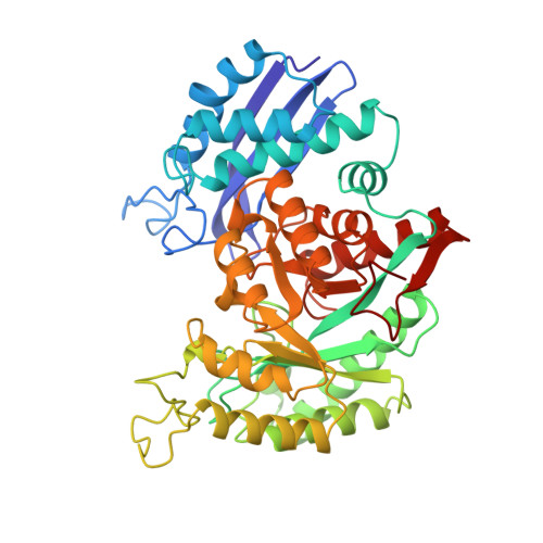
Octahedral coordination at the high-affinity metal site in enolase: crystallographic analysis of the MgII--enzyme complex from yeast at 1.9 A resolution.

Wedekind, J.E.Reed, G.H.Rayment, I.

(1995) Biochemistry 34: 4325-4330

  • DOI: 10.1021/bi00013a022
  • Primary Citation of Related Structures:  
    1EBH

  • PubMed Abstract: 
  • The structure of the Mg2+ complex of yeast enolase has been determined from crystals grown in solutions of poly(ethylene glycol) at pH 8.1. Crystals belong to the space group P2(1) and have unit cell dimensions a = 72.5 A, b = 73.2 A, c = 89.1 A, and beta = 104 ...

    The structure of the Mg2+ complex of yeast enolase has been determined from crystals grown in solutions of poly(ethylene glycol) at pH 8.1. Crystals belong to the space group P2(1) and have unit cell dimensions a = 72.5 A, b = 73.2 A, c = 89.1 A, and beta = 104.4 degrees. There is one dimer in the asymmetric unit. The current crystallographic R-factor is 19.0% for all recorded data to 1.9 A resolution. The electron density indicates a hexacoordinate Mg2+ at the high-affinity cation binding site. The octahedral coordination sphere consists of a meridional arrangement of three carboxylate oxygens from the side chains of Asp 246, Asp 320, and Glu 295, and three well-ordered water molecules. Octahedral coordination is the preferred geometry for alkaline earth metal ions in complexes with oxygen donor groups. In previous crystallographic studies of enolase, Zn2+ and Mg2+ complexes at the high-affinity site were reported to exist in trigonal bipyramidal coordination. This geometry was suggested to enhance the electrophilicity of the metal ion and promote rapid ligand exchange [Lebioda, L., & Stec, B. (1989) J. Am. Chem. Soc. 111, 8511-8513]. The octahedral arrangement of carboxylate and water ligands in the MgII-enolase complex determined here is most consistent with reports of the Mn2+ and Mg2+ coordination complexes of mandelate racemase and muconate lactonizing enzyme. These latter enzymes have alpha/beta-barrel folds comparable to enolase.(ABSTRACT TRUNCATED AT 250 WORDS)


    Related Citations: 
    • Chelation of Ser 39 to Mg++ Latches a Gate at the Active Site of Enolase: Structure of the Bis Mg++ Complex of Yeast Enolase and the Intermediate, Phosphonoacetohydroxamate, at 2.1 Angstroms Resolution
      Wedekind, J.E., Reed, G.H., Rayment, I.
      (1994) Biochemistry 33: 9333

    Organizational Affiliation

    Institute for Enzyme Research, Graduate School, University of Wisconsin, Madison 53705, USA.


Macromolecules
Find similar proteins by:  (by identity cutoff)  |  3D Structure
Entity ID: 1
MoleculeChainsSequence LengthOrganismDetailsImage
ENOLASEA, B436Saccharomyces cerevisiaeMutation(s): 0 
EC: 4.2.1.11
UniProt
Find proteins for P00924 (Saccharomyces cerevisiae (strain ATCC 204508 / S288c))
Explore P00924 
Go to UniProtKB:  P00924
Entity Groups  
Sequence Clusters30% Identity50% Identity70% Identity90% Identity95% Identity100% Identity
UniProt GroupP00924
Protein Feature View
Expand
  • Reference Sequence
Small Molecules
Ligands 2 Unique
IDChainsName / Formula / InChI Key2D Diagram3D Interactions
CL
Query on CL

Download Ideal Coordinates CCD File 
C [auth A],
E [auth B]
CHLORIDE ION
Cl
VEXZGXHMUGYJMC-UHFFFAOYSA-M
 Ligand Interaction
MG
Query on MG

Download Ideal Coordinates CCD File 
D [auth A],
F [auth B]
MAGNESIUM ION
Mg
JLVVSXFLKOJNIY-UHFFFAOYSA-N
 Ligand Interaction
Experimental Data & Validation

Experimental Data

  • Method: X-RAY DIFFRACTION
  • Resolution: 1.90 Å
  • R-Value Observed: 0.190 
  • Space Group: P 1 21 1
Unit Cell:
Length ( Å )Angle ( ˚ )
a = 72.5α = 90
b = 73.2β = 104.4
c = 89.1γ = 90
Software Package:
Software NamePurpose
X-PLORmodel building
TNTrefinement
CHAINrefinement
X-PLORrefinement
X-PLORphasing

Structure Validation

View Full Validation Report


Entry History 

Deposition Data

Revision History  (Full details and data files)

  • Version 1.0: 1995-04-27
    Type: Initial release
  • Version 1.1: 2008-03-03
    Changes: Version format compliance
  • Version 1.2: 2011-07-13
    Changes: Version format compliance
  • Version 1.3: 2017-11-29
    Changes: Derived calculations, Other