1DWR

MYOGLOBIN (HORSE HEART) WILD-TYPE COMPLEXED WITH CO


Experimental Data Snapshot

  • Method: X-RAY DIFFRACTION
  • Resolution: 1.45 Å
  • R-Value Free: 0.255 
  • R-Value Work: 0.211 
  • R-Value Observed: 0.211 

wwPDB Validation   3D Report Full Report


Ligand Structure Quality Assessment 


This is version 1.5 of the entry. See complete history


Literature

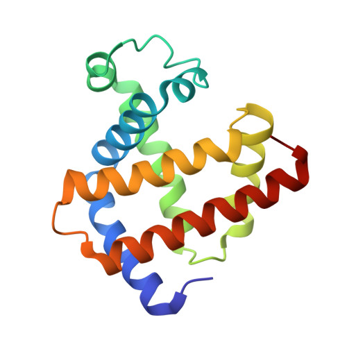
Crystal Structure of a New Ligand Binding Intermediate in Wildtype Carbonmonoxy Myoglobin

Chu, K.Vojtechovsky, J.Mcmahon, B.H.Sweet, R.M.Berendzen, J.Schlichting, I.

(2000) Nature 403: 921

  • DOI: https://doi.org/10.1038/35002641
  • Primary Citation of Related Structures:  
    1DWR, 1DWS, 1DWT

  • PubMed Abstract: 
  • Small molecules such as NO, O2, CO or H2 are important biological ligands that bind to metalloproteins to function crucially in processes such as signal transduction, respiration and catalysis. A key issue for understanding the regulation of reaction mechanisms in these systems is whether ligands gain access to the binding sites through specific channels and docking sites, or by random diffusion through the protein matrix ...

    Small molecules such as NO, O2, CO or H2 are important biological ligands that bind to metalloproteins to function crucially in processes such as signal transduction, respiration and catalysis. A key issue for understanding the regulation of reaction mechanisms in these systems is whether ligands gain access to the binding sites through specific channels and docking sites, or by random diffusion through the protein matrix. A model system for studying this issue is myoglobin, a simple haem protein. Myoglobin has been studied extensively by spectroscopy, crystallography, computation and theory. It serves as an aid to oxygen diffusion but also binds carbon monoxide, a byproduct of endogenous haem catabolism. Molecular dynamics simulations, random mutagenesis and flash photolysis studies indicate that ligand migration occurs through a limited number of pathways involving docking sites. Here we report the 1.4 A resolution crystal structure of a ligand-binding intermediate in carbonmonoxy myoglobin that may have far-reaching implications for understanding the dynamics of ligand binding and catalysis.


    Related Citations: 
    • A Myoglobin Variant with a Polar Substitution in a Conserved Hydrophobic Cluster in the Heme Binding Pocket
      Maurus, R., Overall, C.M., Bogumil, R., Luo, Y., Mauk, A.G., Smith, M., Brayer, G.D.
      (1997) Biochim Biophys Acta 1341: 1
    • Crystal Structure of Photolysed Myoglobin
      Schlichting, I., Berendzen, J., Phillips Jr, G.N., Sweet, R.M.
      (1994) Nature 371: 808
    • Crystallization and Preliminary Diffraction Data for Horse Heart Metmyoglobin
      Sherwood, C., Mauk, A.G., Brayer, G.D.
      (1987) J Mol Biol 193: 227

    Organizational Affiliation

    P-21 Biophysics Group, Los Alamos National Laboratory, New Mexico, 87545, USA. kelvin.chu@uvm.edu


Macromolecules
Find similar proteins by:  (by identity cutoff)  |  3D Structure
Entity ID: 1
MoleculeChainsSequence LengthOrganismDetailsImage
MyoglobinA153Equus caballusMutation(s): 0 
Gene Names: MB
UniProt
Find proteins for P68082 (Equus caballus)
Explore P68082 
Go to UniProtKB:  P68082
Entity Groups  
Sequence Clusters30% Identity50% Identity70% Identity90% Identity95% Identity100% Identity
UniProt GroupP68082
Protein Feature View
Expand
  • Reference Sequence
Small Molecules
Ligands 3 Unique
IDChainsName / Formula / InChI Key2D Diagram3D Interactions
HEM
Query on HEM

Download Ideal Coordinates CCD File 
B [auth A]PROTOPORPHYRIN IX CONTAINING FE
C34 H32 Fe N4 O4
KABFMIBPWCXCRK-RGGAHWMASA-L
 Ligand Interaction
SO4
Query on SO4

Download Ideal Coordinates CCD File 
D [auth A],
E [auth A]
SULFATE ION
O4 S
QAOWNCQODCNURD-UHFFFAOYSA-L
 Ligand Interaction
CMO
Query on CMO

Download Ideal Coordinates CCD File 
C [auth A]CARBON MONOXIDE
C O
UGFAIRIUMAVXCW-UHFFFAOYSA-N
 Ligand Interaction
Experimental Data & Validation

Experimental Data

  • Method: X-RAY DIFFRACTION
  • Resolution: 1.45 Å
  • R-Value Free: 0.255 
  • R-Value Work: 0.211 
  • R-Value Observed: 0.211 
  • Space Group: P 1 21 1
Unit Cell:
Length ( Å )Angle ( ˚ )
a = 63.6α = 90
b = 28.8β = 106.5
c = 35.6γ = 90
Software Package:
Software NamePurpose
X-PLORrefinement
XDSdata reduction
XSCALEdata scaling
X-PLORphasing

Structure Validation

View Full Validation Report


Ligand Structure Quality Assessment 


Entry History 

Deposition Data

Revision History  (Full details and data files)

  • Version 1.0: 2000-03-03
    Type: Initial release
  • Version 1.1: 2011-05-07
    Changes: Version format compliance
  • Version 1.2: 2011-07-13
    Changes: Version format compliance
  • Version 1.3: 2019-05-08
    Changes: Data collection, Experimental preparation, Other
  • Version 1.4: 2019-09-25
    Changes: Data collection, Database references, Other, Source and taxonomy, Structure summary
  • Version 1.5: 2019-11-27
    Changes: Advisory, Derived calculations