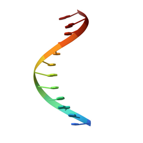
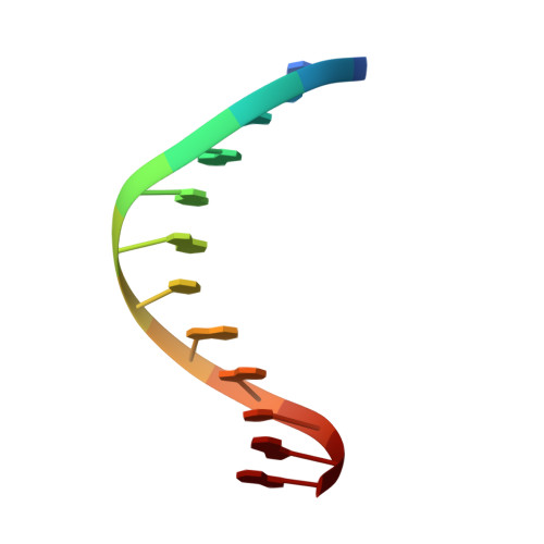
An NMR and molecular modelling analysis of d(CTACTGCTTTAG). d(CTAAAGCAGTAG) reveals that the particular behaviour of TpA steps is related to edge-to-edge contacts of their base-pairs in the major groove
Leporc, S., Mauffret, O., Tevanian, G., Lescot, E., Monnot, M., Fermandjian, S.(1999) Nucleic Acids Res 27: 4759-4767
- PubMed: 10572176 Search on PubMedSearch on PubMed Central
- DOI: https://doi.org/10.1093/nar/27.24.4759
- Primary Citation Related Structures:
1CS2 - PubMed Abstract:
In a previous NMR study we detected the presence of particular motions and hydration properties within the DNA fragment d(CTACTGCTTTAG).d(CTAAAGCAGTAG). Now, we report on an NMR and molecular modelling analysis of this sequence focusing our attention on the biologically important TpA steps. NOe and coupling constant restraints were introduced in three different modelling protocols: X-PLOR and JUMNA used with Flex and AMBER94 as force-fields. Despite their differences the protocols produce similar mean B-DNA structures (r.m.s.d. <1 A). The new information confirms our previous experimental results on the narrowing of the minor groove along the T8T9T10/A17A16A15 run and the sudden widening at the T10pA11 step ending this run. It is further shown that this step displays a large positive roll with its T10:A15 and A11:T14 base-pairs likely stabilised by amino-amino and amino-carbonyl interactions in the major groove. A relationship between roll values and amino-amino and amino-carbonyl distances strongly suggests that electrostatics or bifurcated hydrogen-bonds could be responsible for induction of positive rolls in TpA steps. Such edge-to-edge interactions could explain the slower motions shown by the adenine A15. The influence of these interactions on the stabilisation of particular DNA conformers is discussed using our data and those provided by the recent literature.
- Département de Biologie Structurale, UMR 8532 CNRS, Institut Gustave Roussy, 39-53 rue Camille Desmoulins, 94805 Villejuif, France.
Organizational Affiliation: